U.S. flag

An official website of the United States government

Here’s how you know

Dot gov

Official websites use .gov
A .gov website belongs to an official government organization in the United States.

HTTPS

Secure .gov websites use HTTPS
A lock (LockA locked padlock) or https:// means you have safely connected to the .gov website. Share sensitive information only on official, secure websites.

  • Environmental Topics
  • Laws & Regulations
  • Report a Violation
  • About EPA
Contact Us

Grantee Research Project Results

Final Report: Effects of Nanomaterials on Human Blood Coagulation

EPA Grant Number: R832843
Title: Effects of Nanomaterials on Human Blood Coagulation
Investigators: Perrotta, Peter L. , Gouma, Pelagia-Irene
Institution: West Virginia University , The State University of New York at Stony Brook
EPA Project Officer: Hahn, Intaek
Project Period: February 1, 2006 through January 31, 2009
Project Amount: $375,000
RFA: Exploratory Research: Nanotechnology Research Grants Investigating Environmental and Human Health Effects of Manufactured Nanomaterials: A Joint Research Solicitation - EPA, NSF, NIOSH (2005) RFA Text |  Recipients Lists
Research Category: Nanotechnology , Safer Chemicals

Objective:

Study effects of nanomaterials on the blood coagulation system.

Summary/Accomplishments (Outputs/Outcomes):

1.  Dispersing nanoparticles in biological systems
 
The implications of many nanotoxicology studies are difficult to interpret because investigators often do not adequately document dispersion of nanoparticles used within their experimental system. In collaboration with Dr. Darran Cairns of the Mechanical Engineering Department at WYU, we prepared dispersions of metal oxides down to the l0-20 nm range. Following preparation, the particles remained dispersed when the pH of the media is reduced toward physiologic ranges, and the average particle size did not change over several days as determined by dynamic light scattering (DLS). We found that DLS was the most rapid and reproducible means we had available to document particle size in solution. Other techniques such as atomic force microscopy (AFM) were less reliable because the samples must be dried before analysis. Solution phase or wet AFM was not studied. Nanoparticle dispersion remains a major barrier to studying the effects of nanoparticles in solution.
 
 
2.  Cell interactions with nanostructured hydroxyapatite (HA) particles dispersed on cellulose acetate (CA) Fibers
 
These experiments were carried out in collaboration with Berkeley Lab's Molecular Foundry. Human osteoblasts (SaOS2) cells were used to assess their interactions with Cellulose Acetate/Hydroxyapatite (CA/HA) electrospun mats. l5 mm circular mats in 24-well plates in triplicates were seeded with cells. SaOS2 cells were cultured in Dulbecco's modified Eagle's medium under standard culture conditions. For the cell attachment and proliferation study on plain CA and CA/HA mats, SaOS2 cells were seeded at a density of 50,000 cells/well for MTS assay on the mats and empty wells as controls in triplicates. The cultures were placed at 37oC in a humidified atmosphere containing 5% CO2 for 1 day and 3 days.
 
The Promega Cell Titer 96® Non-Radioactive Assay is used for the metabolic activity and proliferation of SaOS2 cells on CA and CNHA. This assay uses the tetrazolium compound 3-(4,5-dimethylthiazol-2-yl)-5-(3-carboxymethoxyphenyl)-2-(4-sulfophenyl)-2H-tetrazolium; MTS. MTS is chemically reduced by cells into formazan product. Since the production of formazan is proportional to the number of living cells, the intensity of the produced color is a good indication of the viability of the cells. The measurement of the absorbance of the formazan was carried out using 96 well plates following the large volume overnight assay protocol following generation of a standard curve for the MTS assay. Six dilutions (78,500, 15,700, 19,625, 1,570, 157 and 15.7 cells/mL) were prepared from the original 157,000 cells/mL. 1.0 mL of each cell suspension was seeded by transferring the cell dilutions into each well of a 24 well tissue culture plate. Each dilution was performed in three replicates. Then, l 50 µL of the Dye Solution was added to each suspension. The plate was placed at 37oC in a humidified atmosphere containing 5% CO2 in dark. After 4 hours, 1.0 mL of Solubilization/Stop Mix was added to each well. The plate was sealed and kept at 37oC in a humidified atmosphere containing 5% CO2 overnight. 100 µL of the solutions from each well was transferred to a 96 well plate and absorbance was read at 570 nm wavelength and also at 650 nm as a reference wavelength using a Molecular Devices SpectraMax Spectrophotometer.
 
The cells were observed in control samples and on the mats after l day and 3 days (Figure 1). Significantly large differences in the number or growth rate of cells on mats vs. plastic well controls were not observed in these experiments. This indicates that the mats do not have a detrimental effect on cellular growth and proliferation. One might expect to see larger differences in favor of the mats in longer-term experiments once the cells had time to exceed the growth area of the plastic control wells. Because of the low density of fibers, CA mats (consisting of belt-shaped fibers) allowed the cells to pass through them and settle on the plastic bottom of the well during initial seeding. The CA/HA mats had fibrous consistency and retained more cells. Microscopy studies are currently under way to observe the nature of specific cell-HA interactions.
 
Figure 1
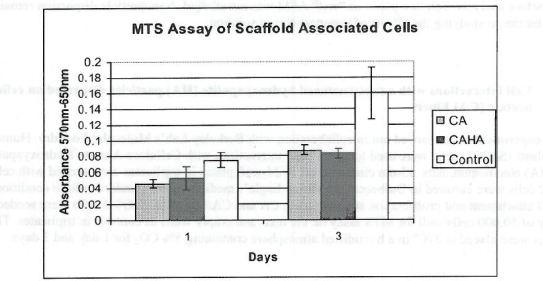
Figure 1. MTS Assay, Absorbance Spectrum after 1 day and 3 days of cells associated with CA and CA/HA mats and Controls. Error bars represent means +- SD
 
 
3.  Nano-chemomechanical transducers
 
A system that transduces chemical energy into mechanical work is called a "“mechanochemical" or "“chemomechanical" system (Okuzaki 1997). The unique feature of this material to trigger large motions in response to change in chemical environment is ideal for use as sensors and actuators. This kind of actuator finds a variety of applications in biology and medicine (e.g., in the detection of biological species) (Gao 2003). Polymer actuators that are chemically stimulated can be used to mimic biological muscles. Porous asymmetric polyaniline membranes have been shown to demonstrate chemical actuation in various solvents such as hexane, ethyl ether, ethyl acetate, tetrahydrofuran (THF), and ethanol. In this material, the chemomechanical actuation is likely caused by the asymmetric structure of the membrane's cross section (Schneider 2007).
 
Cellulose acetate (CA), a natural polymer and a plentiful organic compound found in the higher plants' cell walls (Kennedy 1985), can be easily molded or drawn into fibers for use in various applications. Cellulose based materials allow better control over scaffold design due to their low water solubility (Marques 2005). Cellulose acetate has previously been used as scaffold for functional cardiac cell growth (Entcheva 2004)]. Highly transparent polyaniline-cellulose acetate composite films cast from m-cresol solutions have been reported to possess excellent conductivity and mechanical properties. These films combine the conductivity of polyaniline while retaining the mechanical properties of the host insulating polymer for small content of the conductive polymer in the composite (Pron 1997). Since these composite films possess excellent mechanical properties and are ideal as biocomposite substrates, we investigated the chemomechanical actuation behaviour of these composites for potential application as artificial muscles.
 
Preparation and characterization of polyaniline-cellulose acetate composite films
 
Polyaniline-emeraldine salt (12% w/v) was added to a solution containing cellulose acetate (12% w/v) in acetone. This mixture was sonicated for 2 hours to prevent the precipitation of cellulose acetate. This solvent mixture was cast on a glass substrate and it was allowed to dry at room temperature. The cast film was peeled off from the glass substrate after drying.
 
Setup for measuring chemoactuation
 
A 20 mL beaker containing 10 mL of acetone was sealed to saturate the vapors. Thin strip of the polymer (dimensions 25mm x 4mm) was cut from the cast films. This polymer strip was exposed to the vapor through a small hole. The strip was fixed at one end, leaving the other end free to move (Figure 2). This same setup was used with ethanol replacing acetone. An angular plot was placed behind the beaker to determine the maximum angle of bending.
 
Figure 2
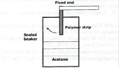
Figure 2. Setup for measuring chemical actuation
 
 
Findings
 
Morphological characterization showed the presence of pores on the surface of the cast films. More pores were found on the surface exposed to the atmosphere compared to those found on the surface in contact with the glass substrate. It is believed that there is a gradient in pore distribution in the casting. This causes the bending of the film. As the film adsorbs on the film, the volume change is higher on the less porous surface, leading to bending towards the more porous surface.
 
When the polymer strips were inserted in a beaker containing acetone, the polymer films were observed to show visible bending. The films were observed to bend to a maximum angle of 40 degrees. The maximum bending angle was attained in 4 seconds. When the seal was broken and the polymer film was found to recover to its original configuration in 3 seconds. This kind of bending-recovery was also observed in ethanol; however the bending was not as pronounced as in acetone vapors. This bending­recovery behavior is believed to be caused by the adsorption and desorption of organic vapors. Figure 3 shows the bending-recovery behavior in acetone. The bending was found to be extensive in acetone and less pronounced in ethanol. Since polyaniline chains are hydrophilic in nature, the bending is dependent on the polarity of the vapor. The higher the polarity of the vapor, the interaction of the polymer chain with the vapor is higher causing extensive bending.
 
Figure 3
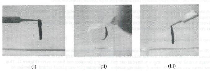
Figure 3. Optical images of bending and recovery in polyaniline-cellulose acetate films (i) Original configureation (ii) Bending in acetome vapors (iii) Recovery in air
 
4.  Conductimetric measurement of cation concentration in solutions using electrospun PANI composites as sensing matrices
 
Studies were conducted to determine the activity of thrombin, the most physiological important clotting protein. Unfortunately, the current technology used to measure thrombin in blood is not ideal because of the relatively low concentrations of thrombin in blood. In addition, the half-life of thrombin is very short. Thus, we are using nanotechnology to develop sensors that are more capable of measuring thrombin in blood. The principle of operation of one such thrombin biosensor involves the conductimetric measurement of NH2+ cations in analyte solutions by electroactive polymers, such as polyaniline (PANI) composites. The first step towards achieving this goal is to demonstrate the H+ concentration (i.e. pH changes) may be monitored accurately and reproducibly by using electrospun cellulose acetate and emeraldine salt (ES) polyaniline composites. The PANI concentration use was 20% and 50% in the electrospun matrix. The films were bonded using sliver paste to gold wires. The distance between the gold electrodes was 2-3 mm. The films were immersed in a solution of 100 mL water with 5 uL of ISA solution to increase and stabilize the pH of the solution to ~10. A Ross pH reference electrode was also immersed in the solution to monitor changes in pH. The resistance of the electrospun composite was measured using a Keithley high resistance multimeter. The pH of the solution was varied by adding 0.1-0.2 mL of acetic acid solutions 0.025%, 0.05%, 0.1%, and 0.2%. It was shown in our work that the 80-20 CA/PANI hybrid is more sensitive to the change in [H+] in the solution than the 50-50 composition. When acetic acid reacts with water the following reaction occurs:
 
Formula
 
Since acetic acid is monoprotic, for every unit of acetic acid, only one H+ is liberated. Water arranges itself around this proton forming an acidic molecule. As the pH of the solution decreases, the concentration of H+ increases in the solution. Above pH 9, polyaniline is deprotonated by the basic solution (considered a proton acceptor) due to the excess CH3COO-, resulting in an increase in the film's resistance. As the pH of the solution decreases the solution becomes a proton donor and the resistance of the film begins to slowly decrease. These studies demonstrated the feasibility of developing such sensors for measuring coagulation proteins like thrombin.
 
 
5.  Preparing bio-composite mats as biosensor substrates
 
The successful incorporation of biological reagents into electrospun nanofibers, and sustained activity through the harsh environment of the electrospinning process has recently been described. Our group has utilized the electrospinning process to create enzymatic biocomposite nanofibers. This experiment for the first time connected the fields of electrospinning and biosensing by employing urease nanofiber mats as novel urea biosensing material. The mechanism used in enzyme-based urea biosensors concentrates on irreversible hydrolysis of urea into ammonia and carbon dioxide in the presence of urease. Blends of polyvinylpyrrolidone (PVP) and urease were electrospun, and formed beaded nanofibers with diameters of 7 to 100 nm. The encapsulated enzyme activity was comparable to its pure form. Further studies showed that solvent substitution and increased polymer concentration allowed for a larger amount of enzyme to be electrospun to form smoother fibers without beads. The successful incorporation of urease into comparable polymer nanofibers demonstrates that this novel design can be used to produce biosensors.
 
 
6.  Hemolytic Properties of Nanoparticles
 
Our laboratory was part of a collaborative project involved in analyzing the hemolytic properties of nanoparticles. This method tests the effect of nanoparticulate materials on the integrity of red blood cells. This test method used diluted whole blood incubated with nanoparticulate material and the hemoglobin released from damaged cells is determined by a quantitative colorimetric assay which included standards. These studies demonstrated the difficulty in standardizing tests of nanotoxicity. Despite standardized procedures and reagents, considerable differences were seen in tests performed at different labs. A major limitation was the difficulty in using blood samples collected at a central site and distributed across the country to different laboratories.
 
 
7.  Electrochemical detection of R-NH2+ as product of reaction of Thrombin on its substrate
 
Additional work was performed to develop a nanosensor designed to detect low levels of thrombin as discussed above. This sensor detects the thrombin-catalyzed hydrolysis of a specific thrombin substrate (Benzoyl-Phe-Val-Arg-AMC, HCl) by using a conducting polymer that measures changes in electrical resistance in the presence of ions. Such a sensor would be useful in measuring small changes in thrombin that occur in a variety of human diseases, both in the blood, and at the surface of cells and blood vessels.
 
Thrombin (1KU) was reconstituted in 50 mM NaCl. 100 units of thrombin were dissolved in 1 mL of NaCl solution. Thrombin substrate was reconstituted in distilled water. 1 mM solution of thrombin substrate was made. Ammonia electrodes were used for -NH2+ detection. 1 mL of thrombin solution (100 units) was added to 1 ml of 1 mM solution of peptide and electrode potential was noted after 2 minutes of reaction time. Similarly, readings were taken at 5 minutes and 10 minutes. It was seen that electrode potential decreases as reaction time increases indication more and more cone of -NH2+ species as reaction time increases and also proves that thrombin acted on its substrate (Figure 4).
 
Figure 4
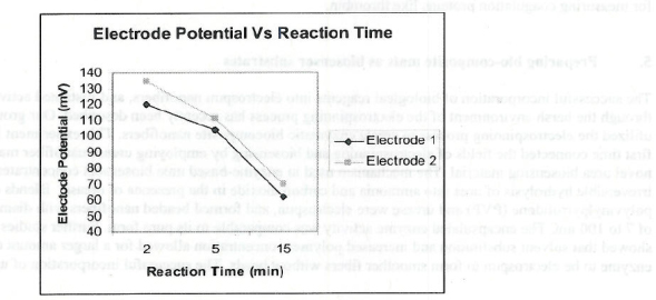
Figure 4. Plot of electrode potential (mV) Va Reaction time (Min). Two sets of readings by two electrodes are shown with similar trends and are in good agreement
 
 
8.  Effects of inhaled nanoparticles on coagulation
 
We collaborated with Dr. Timothy Nurkewicz at WVU, who is using an inhalation model to study the vascular system of rats exposed to particles in the nanosized range. Blood is collected from the jugular vein to minimize clotting protein and platelet activation from control and exposed rats into sodium citrate (0.105M, 3.2%) as the anticoagulant. This is important because blood is usually collected into EDTA, which is not useful for clotting studies. Platelet-poor plasma was removed using standard centrifugation protocol at 4°C and aliquots were stored at -80°C until use.
 
The rationale for performing coagulation studies is that common human diseases including myocardial infarction and stroke are caused by abnormalities of blood coagulation that predispose to thrombosis (clots). These diseases are influenced by environmental factors, however, not all risk factors for clotting disorders are known. Nanomaterials that enter the workplace could enter the body through inhalation, and these exposures could alter the blood coagulation system. Although in vitro studies of the effects of nanosized materials on the blood coagulation proteins can be performed, in vivo studies are needed because effects on blood clotting cannot be predicted with certainty using by in vitro experiments alone.
 
Coagulation studies were performed on rats exposed to various doses and sizes of titanium dioxide (TiO2) particles through inhalation. Doses of TiO2 ranged from 6 to 100 ug (0.015-0.1) during the 24 hour exposures. Whole blood was drawn from control and test animals into sodium citrate anticoagulant (0.105M, 3.2%, 1:10 ratio citrate to whole blood). Platelet-poor plasma was prepared by centrifugation at 4°C and stored at -80°C until use. Plasma prepared in this way was used for all studies described in this section.
 
Endogenous thrombin potential (ETP) of the rat plasma was measured using a kit (Technothrombin TGA, Columbus, OH) containing a phospholipid micelle reagent (TGA RC High Reagent) and a fluorogenic thrombin substrate (Z-GR-AMC). Plasma (40 uL) diluted with buffer was added to black bottom 96-well microplates, after which the TGA reagent and TGA substrate (50 uL) were added. Fluorometric measurements were made at 60 sec intervals for up to l hour using a kinetic plate reader (Beckman DTX 880- Beckman Coulter, Fullerton, CA) pre-warmed to 37°C and set to appropriate wavelengths (excitation 390 nm, emission 465 nm). A thrombin calibration curve was generated using known concentration s of thrombin according to the kit protocol in order to quantify thrombin generation. The intra-assay and inter­assay variability of the ETP assay for thrombin generation were measured at 9% and 6%, respectively.
 
Fibrinogen (FBG) plays a major role in coagulation, and both elevated and decreased levels have clinical significance. Elevated plasma FBG has been identified as an independent risk factor for coronary atherosclerosis and ischemic heart disease. Individuals with congenital absence of FBG, termed afibrinogenemia, have prolonged bleeding times. A Micro-plate Colorimetric ELISA Method was used to measure Fibrinogen levels in rat plasma samples. Fibrinogen Standard Curve was generated according to the protocol which facilitated the quantification of fibrinogen level in the samples. These studies are ongoing.
 
A customized kit (LINCOplex Rat CVD Panel, Millipore, St. Charles, MO) was used to quantify specific analytes in rat plasma including fibrinogen, von Willebrand factor (vWF), troponin 1 and troponin T. This technology allows multiplexed detection of proteins from a single sample. This 96 well microplate immunoassay utilizes distinct color microsphere beads coated with capture antibody. Sample is added to these beads and the analyte is captured, after which a fluorescent tagged detection antibody is then added. The Luminex analyzer lasers detect both bead dyes and tagged detection antibody. Analyte concentrations are calculated using the built-in software. Fibrinogen was also quantified in rat plasma using an ELISA assay (AssayMax Rat Fibrinogen, AssayPro, St. Charles, MO) according to manufacturer's instructions.
 
The endogenous thrombin potential (ETP) assay was used to measure the ability of rat plasma to generate thrombin, an assay that utilizes a fluorogenic thrombin substrate. The total amount of thrombin generated over the duration of the readings is determined by measuring the area under the curve (AUC). The maximum amount of thrombin generated (peak thrombin generation) at any point in time is also determined. Thrombin generation was significantly higher in rats exposed to either ultrafine or fine particles at the 10 ug dose than in control animals (p < 0.05) (Table 1). The effect was more pronounced for the fine particle exposure.
  Dose Thrombin generation (nM/min) Peak thrombin (nM)
Control (n=17) NA 2972 (708) 195 (45)
Ultrafine (n=13) 0.03 (10) 3544 (332)* 244 (24)*
Fine (n=8) 0.03 (10) 4277 (156)*+ 273 (13)*+

Table 1 Endogeneous thombin potential (ETP) of plasma obtained from rats exposed to
and ultrafine TiO2 as compared to controls. Values denote means (standard deviations).

* Denotes statistically significant differences between the exposed and control group.

+ Denotes statistically significant differences between the exposed groups. 

Total thrombin generation was studied at various doses for the fine particle exposures over 24 hours. ETP was significantly higher in all dosing groups studied as compared to controls (Table 2). No dose-response relationship could be seen between exposure doses and ETP as an increased effect was not seen as exposure increased. The most pronounced effect on thrombin generation occurred in the 10 ug group.
Dose N Thrombin generation (nM/min)
Control 15 2890 (705)
0.015 (6) 5 3824 (285)*
0.0. (10) 8 4277 (182)*
0.06 (19) 9 3696 (354)*
0.10 (67) 11 3240 (791)*
0.15 (90) 5 3692 (264)*

Table 2. Endogenous thrombin potential of plasma in rats exposed to various doses of 
fine TiO2 particles for 24 hours by inhalation. Values denote means (standard deviations)

* Denotes statistically significant difference between the exposed and control group.

 
Using the Luminex assay, very few differences were seen between fibrinogen and vWF levels between exposed groups and controls (Table 3). This is partly explained by the variability of these assays, especially the fibrinogen assay. The exaggerated increase in fibrinogen in the 90 ug fine group was explained by very high values in approximately two-thirds of the animals in this group. Other animals in this group had values that were more consistent with other groups. Using the rat fibrinogen ELISA assay, no significant differences could be found between any of the groups (Table 4). For vWF measurements, only the 10 ug ultrafine exposed group had values significantly higher than controls (Table 3).
Particle Dose Fibrinogen vWF
Fine 0.10 (67) 109,233) 140 (92)
Fine 0.15(90) 1,184692 (630, 111)* 155 (95)
Control NA 197,544 (30 1, 868) 105 (58)
Ultrafine 0.015 (6) 139,740 (187,621) 135 (69)
Ultrafine 0.03 (10) 81,810 (13,951) 1 85 (130)+
Ultrafine 0.06 99,672 (1 06, 580) 99 (65)
Ultrafine 0.15 (38) 52,943 (44,247) 138 (109)

Table 3. Fibriogen and vWF of plasma in rats exposed to various doses of fine and ultrafine
Ti02 particles for 24 hours using a Luminex assay. Values denote means (standard deviations)
flourescent units. 

* Denotes statistically significant differences between this group and all other exposed groups and contros (p>0.001).
+ Denotes statistically significant differences between this group and control (p<0.01).

 
Particle Dose Fibrinogen (ug/mL)
Fine 0.10 (67) 20 (12)
Fine 0.15 (90) 87 (8)
Control NA 51 (24)
Ultrafine 0.03 (10) 65 (26)
Ultrafine 0.06 (19) 68 (27)

Table 4. Fibrinogen and vWF of plasma in rats exposed to vaiours doses of fine and ultrafine
T0)2 particles for 24 hours using a rat fibrinogen ELISA assay. Values denote means (standard deviations)

 
Significant elevations were demonstrated in both troponin I and troponin T at a single dose in the fine and ultrafine exposure groups (fine 67 ug and ultrafine 6 ug) (Table 5). Although these differences were statistically significant, it is unclear if they truly reflect myocardial injury. There were no dose-response effects in either troponin I or troponin T in the exposed animals.
 
Particle Dose Troponin I Troponin T
Fine 0.10 (67) 73,612 (4,150)* 7.042 (1070)*
Fine 0.15 (90) 67,793 (3,895) 5,0.8 (975)
Control NA 69,495 (2.801) 61.178 (1704)
Ultrafine 0.15 (6) 75.026 (5,237)* 7,059 (404)*
Ultrafine 0.03 (19) 670,1 (4,4017) 6,379 (617)
Ultrafine 0.06 (19) 68,705 (3,446) 6,467 (350)
Ultrafine 0.15 (38) 7 1.639 (3,519) 6.594 (779)

Table 5. Troponin T and Tropinin I levels in plasma of rats exposed to various doses
of cine and ultrafine Ti02 particles for 24 hours using a LUminex assay. Values denote
mean (standard deviations) flourescent units.
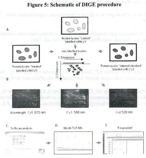
 
Discussion
 
We studied plasma obtained from rats exposed to ultrafine and nanometer-sized particles through inhalation. The endogenous thrombin potential (ETP) assay was utilized because of the critical role thrombin plays in blood clotting. Thrombin catalyzes the conversion of fibrinogen, which circulates in blood in a soluble form, to a fibrin clot. The ETP estimates the ability of plasma to generate thrombin, and has clinical relevance in terms of determining risk of clot formation (Baglin 2005). For example, individuals with certain diseases form clots because of excess thrombin generation. Differences in other important clotting proteins including fibrinogen and von Willebrand factor (vWF) were also determined between exposed and control animals.
 
We found variable changes in coagulation parameters for rats exposed to fine and ultrafine TiO2 particles. The most consistent of these changes were seen when ETP was measured in rats exposed to the 10 ug doses of fine and ultrafine particles. Those results suggest that these exposures favor increased thrombin generation, which has been associated with an increased ability to form clots (Tappenden 2007). Other variable changes were seen for fibrinogen, which has been shown through clinical, experimental, and epidemiologic studies to be associated with a risk of clotting (Doutremepuich 1998). No dose-response relationships were seen for any of the coagulation parameters studied.
 
Previous studies of blood coagulation parameters in rats exposed to ambient particulate matter have had similar difficulties demonstrating consistent exposure-related effects (Nadziejko 2002). As in our studies, several statistically significant differences in clotting parameters have been described; however, these changes cannot be clearly labeled as "adverse effects." Variable increases in clotting proteins that increase in concentration during inflammatory reactions such as vWF and fibrinogen observed in our studies have been described in other animal models studying the short-term inhalation effects of ultrafine carbon black particles (Gilmour 2004). It is unclear if these studies translate to the human clotting system. All studies performed to date have utilized healthy rats. Humans who are exposed to particles through inhalation are in various states of health and may already have risk factors for clotting abnom1alities. It is unclear how these individuals will respond to short and/or long-term inhalation exposure.
 
 
9.  Correlating changes in coagulation with cardiac marker status and inflammatory markers (Luminex Assays)
 
Inflammatory mechanisms play a vital role in the initiation, maintenance and progression of vascular diseases leading to coronary artery disease. Rat Multiplex panels (Millipore Corp) are available for the study of the pathogenesis of CVD. These panels are designed for the simultaneous analysis of multiple cardiac biomarkers reflecting oxidative stress, inflammation and vascular or cardiac integrity. Analytes tested include IL-6, MCP-1, MPO, TNF-alpha and VEGF. We had difficulty obtaining reproducible results for inflammatory markers utilizing Luminex technology. This may have been related to technical problems or limitations of the assay for measuring lower levels of cytokines.
 
 
10.  Proteomic studies
 
The effects of inhaled fine (~1000 nm diameter) and ultrafine (~123 nm) TiO2 on rat plasma proteins were studied utilizing differential in gel electrophoresis (DIGE) (Figure 5). Plasma samples were collected animals exposed to the TiO2 aerosols in controlled chambers at concentrations that do not alter bronchoalveolar lavage markers of pulmonary inflammation or lung damage. Aerosol concentrations (mg/m3) studied included 0.15 and 0.03 for ultrafine and 0.10 for fine particles [Nurkiewicz 2008]. Plasma proteins were solubilized in a cell lysis buffer and three cyanine dyes (Cy2, Cy3, Cy5) were used to label the proteins of the test and control groups, as well as a mixture of the two which served as an internal standard (Cy2) (Table 6).
 
Figure 5
Sample Cy2 Cy3 Cy5
1 Internal Standard Control 0.15
2 Internal Standard Control 0.03
3 Internal Standard Control 0.1
4 Internal Standard 0.15 0.1
5 Internal Standard 0.03 0.1
6 Internal Standard 0.03 0.15

Table 6. Experimental design

 
Mixtures of labeled proteins were separated by 2D gel electrophoresis. First dimension isoelectric focusing was performed on 24 cm Immobiline DryStrips with a nonlinear pH 3-10 gradient using an Ettan IPGphor (Amersham Biosciences) at 20°C. The strips were rehydrated for 13 hrs in rehydration buffer containing a mix of Cy2-, Cy3- and Cy5-labeled proteins. Isoelectric focusing was performed in three steps: at 300 V for 1 hr, at 1000V for 1 hr, and at 8000V for 3 hr. Prior to second dimension, strips with separated proteins were equilibrated, reduced, and separated in the second dimension by SOS-PAGE on I 0% polyacrylamide gels. The separation was performed using an Ettan DALTsix unit (Amersham Biosciences) at 25 mA/gel constant current until the dye front migrated out of the gel. The fluorescently labeled proteins were then visualized and quantified using a Typhoon 9400 Variable Mode Imager (Amersham Biosciences). Protein amounts in the test and control samples were quantified based on their corresponding fluorescence intensities, and their molar ratios and volumes were calculated using SameSpots proteome expression analysis software (Progenesis). Separated proteins were identified by excising spots from Comassie-stained 2D gels and digesting with trypsin using standard protocols. Peptides in each tryptic digest were separated and characterized by MALDI-TOF-M S, as well as LC/MS/MS and database searching. MALDI-TOF-MS was operated in the positive ion mode measuring masses from m/z 800 to 3000. Data files were searched against the IPI Rat database (v3.46) matching 5 or more peptides with mass error less than 50 ppm. LC/MS/MS was operated in the positive ion mode measuring masses from m/z 400 to 1500. The Bioworks Browser (v3.2) was used to search raw data files against the IPI Rat database (v3.46). The lysates to be analyzed are labeled with fluorescent dyes (Cy3 and Cy5), while an internal standard is labeled with Cy2. (a). The labeled samples are combined and separated according to pH (1st dimension) and molecular weight (2nd dimension) in the gels (b). Proteome expression analysis (c). All differentially expressed proteins, which were up- or down-regulated with a p value of < 0.05 were picked and measured via MALDI-TOF-MS and LC/MS/MS (d). Peptide mass fingerprint analysis with the Bioworks browser (Corzett 2006).
 
Overall, 428 distinct protein "spots" were identified by 2 dimensional gel electrophoresis. Of these, 72 spots were quantitatively different (Figure 6) between the test groups and controls (p < 0.05). Characterization of these proteins by mass spectrometry (MALDI-TOF or LC/MS/MS) revealed approximately 35 distinct proteins (Table 7). The biological functions of many of the proteins identified included roles in acute phase responses, inflammation, and blood coagulation. The functions of a number of the proteins identified were either poorly understood or not previously implicated in toxic responses.
 
We concluded that exposure to fine and ultrafine TiO2 through inhalation causes significant changes in the rat plasma proteome. These changes may be directly involved in the potential adverse effects of particle exposure, or may serve as markers of toxicity. Additional studies are needed to determine the specific proten "pathways" involved in the adverse health effects of small particle exposure. Proteomic profiling techniques are effective in detecting changes in plasma proteins following environmental/occupational exposures, and could help identify "biomarkers" of particle toxicity.
 
Figure 6
 
Table 7
 
 
11.  Assessing the toxicological properties of nanostructured hydroxyapatite clusters embedded in polymer nanofibrous mats seeded with osteoblasts
 
This reported work is a novel approach to nanotoxicology and tissue/organ scaffolding and it studies how biomaterials' properties of polymer hybrids affect their interactions with living cells. It focuses on assessing the toxicological properties of osteinductive biomaterials, in particular nanostructured Hydroxyapatite (HA). Nanostmctured bioceramics belong to a class of important functional inorganic materials used in health applications and cosmetic products. Human bone is composed of nano-assembled collagen type I and hydroxyapatite (Lee 2007). So HA has been widely studied in bone tissue engineering as bioactive and biocompatible inorganic materials. Many studies have shown that organic and inorganic composite scaffolds support attachment, differentiation and proliferation of osteoblasts. The hybrids of bioceramics with polymers comprise the new generation of biosensors, filtration devices, etc., and thus it is important to understand how they behave when interfacing with living cells and organisms. It takes materials science expertise to fully characterize the structural and chemical changes that materials experience upon interactions with the cells and their environment. Cellulose Acetate (CA) has been widely used in various applications, such as in dialysis membranes, in vitro hollow fibers perfusion systems, as surfaces for cell expansion and guided bone regeneration membranes (Entcheva 2004). CA has low water solubility, good biocompatibility, supporting and guiding structures for cell culture and excellent shape conforming properties (Martson 1999) which make CA an excellent candidate for bone tissue scaffolds. Composites of CA and HA thus are envisioned to be important materials for regenerative medicine and these may be nanomanufactured for use in biomedical applications. Nanocomposites of CA/HA are studied here to assess their toxicological properties. CA and HA solutions were electrospun together to produce nanofibrous scaffold structures in a single step. Cellulose Acetate (CA) and CA/HA Cellulose Acetate/Hydroxyapatite (9.23%) scaffolds were set up in 24 well plates; seeded with SaOS-2 cells and incubated for 1, 7 and 14 days. Cell viability and proliferation were assessed by MTS and Picogreen assays. Cell differentiation and functionality were evaluated by ALP, osteocalcin and Von Kossa procedures. Samples were also collected for electron microscopy to examine cell spreading and morphology. The studied nanocomposites show good in-vitro performance with respect to the growth and proliferation of osteoblast cells and they may be safely used in bone regeneration applications.
 
Materials and Methods:
 
HA powder was prepared from raw eggshells calcinated at 900°C. Short duration (30 min) heat treatment of eggshells caused their color change from white to black, thereafter; they became white after 3 h heating. The color change suggests that most of the organic materials were removed. Powders were then crushed in an agate mortar and reacted with phosphoric acid powder (50:50 wt % H3P04) in an exothermic reaction. Products were milled in ethanol solvent for 10 h by ball mill, after which polyethylene glycol (PEG) was added to portions of the powder. Batches were sieved with 100 µm mesh and green compacts were formed through dry pressing at 220 MPa. HA samples were then sintered in air at 900°C for 2 h while limiting the heating rate to 2°C/min. The structural and morphological evolution of HA particles has been detailed (Balazsi 2009). The sintered HA was crushed to powder form in order to introduce HA into the CA solution.
 
Cellulose acetate powder with an acetyl content of 40% (Fluka Chemie CH-9471, Buchs, Switzerland) was mixed with acetone at room temperature and sonicated for 1 h to form a 15 wt/vol% solution for electrospinning. CA powder was mixed with acetone at room temperature to form a 15 wt/vol% solution and 9.38% w/v HA(hydroxyapatite) nanoparticles was added to acetic acid. This mixture was combined to prepare a solution containing 71-29 vol% Acetone/Acetic acid, respectively. This mixture was sonicated for I hour before electrospinning.
 
Electrospinning conditions: The electrospinning apparatus consists of a high-voltage power supply that provides up to 40 kV, two electrodes, a metering pump, a glass syringe attached to a 22 gauge conducting needle and an aluminum foil collector. One of the two electrodes is attached to the syringe need le and the other is attached to the collector. For electrospinning parameters, a flow rate of 9.6 mL/hr and voltage of 19 kV were used. The distance from the collector to the needle was 10 cm.
 
Cell Culture and Conditions: Scaffolds were seeded into wells of a 24-well tissue culture plates. Stainless steel retaining rings (snap rings) (McMaster-Carr #9158OA 161) were used to secure the scaffolds in the bottom of the wells. Scaffolds were sterilized by incubating with 70% ethanol for 30 min. The ethanol was removed and the scaffolds were washed twice with sterile water. The wells were then filled with Dulbecco's modified Eagle's medium containing 10% fetal bovine serum (FBS), 1% L-glutamine and 1% antibiotic/antimycotic (DMEM complete medium) [All from Sigma] and allowed to equilibrate overnight. Human osteoblast-like cells (SaOS2) were grown at 37oC, 5% CO2 in a humidified incubator in DMEM complete medium
 
The cells were trypsinized with 0.25% trypsin- EDTA (Sigma) and seeded on the scaffolds at a density of 50,000 cells per well in a volume of 1.5 ml. Samples were set up in triplicate wells. On day 3 the medium was changed to DMEM complete medium supplemented osteogenic factors 50 ug/mL ascorbic acid, 10 mM ß-glycerolphosphate and 10-8 M dexamethasone (All from Sigma). The cells were cultured for up to 14 days and the medium was changed every 3 days. (On days 8 and 9, there was a problem with the CO2 supply to the incubators and the cells were without CO2 for ~ 2 days. This may have impacted the cells collected for the day 14 time point). MTS Assay The CellTiter 96® AQueous Assay (MTS Assay-Promega #G3580) uses the novel tetrazolium compound (3-(4,5-d imethylthiazol-2-yl)-5-(3-carboxymethoxyphenyl)-2-(4-sulfophenyl)-2H-tetrazolium, inner salt; MTS) and the electron coupling reagent, phenazine methosulfate (PMS). MTS is chemically reduced by cells into formazan, which is soluble in tissue culture medium. The measurement of the absorbance of the formazan can be carried out using 96 well microplates at 492 nm. The assay measures dehydrogenase enzyme activity found in metabolically active cells (cell viability).
 
After cells were cultured for 1, 7 and 14 days, the scaffolds were transferred to fresh wells and all wells were rinsed with PBS IX. The PBS was aspirated and 150 uL of fresh medium was added to each well. Cell titer reagent (30 uL) was added to each well and the plates were returned to the incubator for 2 hours. A 100 uL aliquot of each sample was transferred to wells of a 96 well plate (including a media only blank). The absorbance at 490nm was recorded using a 96 well plate reader. For quantitation, a standard curve was prepared using SaOS-2 cells ranging from 1,000-150,000 cells per well.
 
Picogreen Assay Quant-iT™ PicoGreen® dsDNA reagent (Invitrogen # P7581) is an ultra-sensitive fluorescent nucleic acid stain for quantitating double-stranded DNA (dsDNA) in solution (quantitation of relative cell number). After cells were cultured for 1, 7 and 14 days, the scaffolds were transferred to fresh wells and all wells were rinsed with PBS 2X. 300 uL of TE buffer was added to each well and samples were freeze-thawed 3 times to release the DNA from the cells. For quantification, a standard curve was prepared with DNA in range of 1-1000 ng/ml. Background signal was evaluated using the scaffolds without cells. A 100 uL aliquot of each sample was mixed with 100 uL of working reagent and incubated for 5 min. at room temperature. The samples were read on a fluorescence plate reader with an excitation of 488 nm and emission of 520 nm.
 
Alkaline Phosphatase Detection: Alkaline Phosphatase (ALP) is an osteoblast phenotypic marker. After cells were cultured for 1, 7 and 14 days, the scaffolds were transferred to fresh wells and all wells were rinsed with PBS 2X. Samples were lysed in 200 u L CellLytic M (Sigma). The samples were assayed using the Alkaline Phosphatase Detection K it (Sigma #APF). A 20 uL aliquot of each sample was mixed with 180 uL of assay mixture and incubated for 2 hours in the dark. The samples were read on a fluorescence plate reader at an excitation of 360 nm and emission of 440 nm. To compare the ALP on a per cell basis, ALP activity was normalized, by dividing the number obtained by protein concentration of the cell lysates (Pierce - BCA assay).
 
Osteocalcin Detection: Osteocalcin is a marker of an osteoblast phenotype. To evaluate the osteocalcin secretion levels, 24 hour media collections were taken at day 1, 7 and 14 and tested in the Osteocalcin Instant ELISA (Bender MedSystems #BMS2020TNST). To minimize background levels of osteocalcin from FBS, media containing 2% FBS was used for the 24 hour collections. Samples were assayed in duplicate and compared to a standard curve.
 
Von Kossa Staining - Mineralization: This technique is for demonstrating deposits of calcium or calcium salt so it is not specific for the calcium ion itself. Samples were fixed with 100% ethanol for 30 min. and rinsed with water. Samples were treated with a solution of 1% Silver Nitrate and incubated under a UV light for 20 min. The wells were washed with water and de-stained with 5% Sodium thiosulfate for 2 min.
 
Cell Morphology - Electron Microscopy (EM): To evaluate cell morphology on the scaffolds samples were prepped for EM staining. Samples were washed 2 times with PBS and fixed with 2.5% glutaraldehyde (Sigma) for 2 hours then washed with PBS. The cell scaffold constructs were then attached to aluminum stubs, sputter-coated with gold, and then examined under a LEO Gemini Schottky FEG scanning electron microscope. The working distance used for this study was varied between 8-10- mm and the operating voltage was varied from 5 to 20 kV. The fiber diameter, pore diameter, and cell diameter data were determined using Image J software.
 
Results:
 
Viability and Proliferation: The effects of the scaffolds on cell viability and proliferation were evaluated by the MTS and Picogreen assays, respectively. In the viability assay, MTS is converted to formazan by metabolically active cells. The relative cell numbers were measured using the Fluorometric double-Stranded DNA quantitation kit, Picogreen. There was a significant increase on both cell viability and number from day 1 to day 7 for the control wells (cells on plastic). Both of the scaffolds, CA and CA/HA, had dramatically lower levels of cell viability and cell number compared to the plastic controls. Cells grown on the scaffolds exhibited little to no growth between day 1 and 7, but appear to show a trend toward cell growth by the day 14 time point. (Figs. 7 and 8). There was a decrease in viability and proliferation for the day 14 control wells. This result is possibly due to the CO2 problem with the incubator.
 
Phenotype retention: ALP activity is a marker of an osteogenic phenotype. All of the samples showed an increase in signal from day 1 to day I 4. The cells grown on the CA and CA/HA scaffold had lower levels of ALP than the cells grown on plastic. When comparing the CA to CA/HA scaffolds, the ALP activity levels were of a similar magnitude. (Figure 9). Osteocalcin is also marker of an osteoblast phenotype. None of the samples showed signal above that seen with the negative control, media + 2% FBS. There is some evidence in the literature that the osteocalcin production of static cultures is low to none and that perfusion cultures are required to induce this marker in cell culture.
 
Figure 7
 
Figure 8
 
Figure 9
 
Mineralization: The detection of mineralization was performed on days 1, 7 and 14. Staining was also done on scaffolds without cells to evaluate the background staining. The scaffolds without cells showed only a slight background staining even though some of the scaffolds contained hydroxyapatite which contains calcium. There was a clear increase in mineralization for all of the samples between day 1 and day 14. The CA/HA showed the most intense staining at each time point with the day 14 samples being the most intense (Fig 10). One observation made during the staining was that during the washes and staining procedures, some of the mineralization that was observed in the control samples appeared to be washing away.
 
Electron Microscopy Imaging of the Scaffolds: Figures 11-13 present the morphology and structure of the CA, CA/HA, and their comparison, respectively. For CA scaffolds, the average diameters of cells for 1, 7 and 14 days culture are 15.14 µm, 17.33 µm and 26.47 µm respectively. For CA/HA composites, the amount of cells is greater than CA scaffolds in each culture period and the cell size is larger than the ones on pure CA scaffolds. The average diameters are 14.77µm, 29.47 µm and 31. 17 µm for 1, 7, 14 days culture. The as-received electrospun scaffolds showed uniform distribution, porosity and ultrafine fiber diameters (Fig. 14). Pure CA scaffolds have a larger fiber size at diameter of 2.9 µm on average. By contrast, due to lower polymer concentration, the CNHA composite electrospun fibers have a 300 nm average diameter. The average size of hydroxyapatite nanoclusters on the fibers is 35 nm (Fig. 15). The average pore diameter in the CA scaffolds is about 5.82 µm and in CA/HA scaffolds it is about 3.19 µm. The relative porosity of the as-spun scaffolds is 43% for the CA and 30% for the CA/HA based on calculations within a layer.
 
Figure 10
 
Figure 11
 
Fogire 12
 
Fogure 13
 
Fogure14
 
Fogure 15
 

Conclusions:

Discussion:
 
There are several key issues addressed in this report regarding the nature of cell-materials interactions:
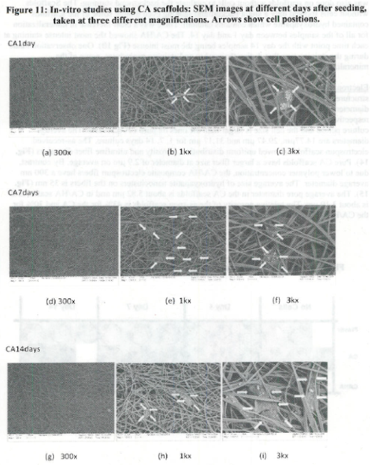
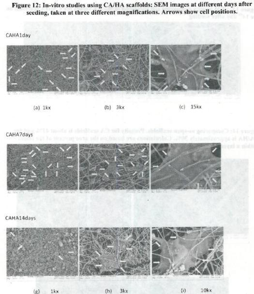
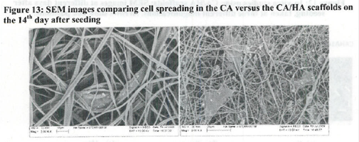
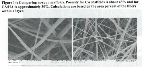
  • The use of open nanomanufactured scaffolds (porosity effects)
  • Relationship between adhesion to nanostructured materials/cues vs cell differentiation
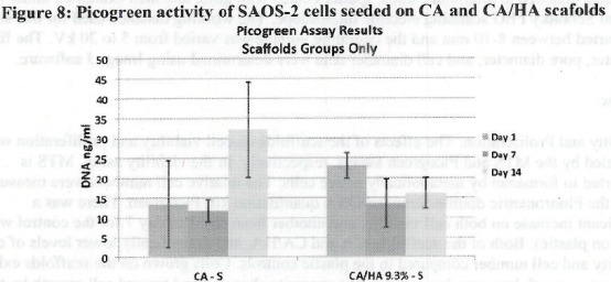
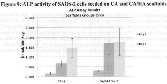
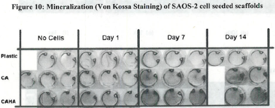
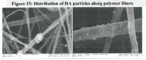
  • Observed pattern of mineralization on scaffolds
The work described in this report is based on the use of uniformly dispersed nanohydroxyapatite clusters on the surface of fibers in non-woven mats of biocompatible polymers. The choice of anchoring bone cells (osteoblasts) in this study was meant to establish their affinity to nano HA as opposed to nano CA. Cellulose and its derivatives, though biocompatible, are not osseoinductive materials, and osteoblasts were not expected to interact strongly with them. Evidence for this fact is given by the micrographs in Figures 11 and 12. Cells would grow only between two CA fibers and along their length in a "panel-looking" configuration in the pure polymeric scaffolds, whereas they were seen forming elongated, widely-spread, complex, “tent­-like” networks involving multiple fibers at different layers, using the HA nanoclusters as anchoring sites in the hybrid mats.
 
Furthermore, all evidence of mineralization on the CA scaffolds is on the surface of the cells (see white spots on cell in Figure 11c), away from the fibers, whereas the hybrids exhibit mineral formation near anchoring sites, presumably at the interfaces with HA. This is an important result which deserves further attention as earlier Raman microprobe studies by other workers [Walters 1991] on HA implants-bone interfaces, as developed in animal implantation studies, have revealed the presence of mixed phosphate mineral phases at such interfaces. It is unclear how the change from stoichiometric HA to the mineral matrix of bone occurs.
 
It is known that nanostructured ceramics promote cell growth (osteoblasts in particular) (Webster 2000), even though it is not clear by what mechanism this is occurring, i.e. via protein adhesion and controlled release or by other means (Habibovic 2007). There is a need for further studies, animal studies in particular, and post mortem histochemical studies need to be performed on the implanted mats to chemically and biologically evaluate the differences in the fiber-cell vs bioceramic-cell interfaces. At the same time, there has been preliminary data supporting the use of open scaffolds in bone engineering (Scaglione 2009), as high porosity scaffolds in in-vivo animal studies appear to promote vascularization of scaffolds thus enhancing cell viability and proliferation, even though this was not always obvious in in-vitro studies carried by the same workers (Hutmacher 2007).
 
Finally, based on a recently published work (Oh 2008) involving stem cells and nanostructured scaffolds, the higher the adhesion of a cell to a nanoentity, the lower its potential to differentiate to the intended functionality. CA scaffolds have cells strongly adhering to individual fibers. CA/HA scaffolds had few adhesion points but an overall free surface separate from the scaffold. Although the assays used here have confirmed the retention of bone phenotype for the cultured cells on both scaffolds, there was no quantitative measurement of their relative degree of differentiation, primarily due to the presence of CA containing HA in the hybrid mats. The elongated shape of the cells on CA/HA scaffolds, though, suggests that there are more likely to have maintained their functionality.
 
In summary, the CA and CA/HA scaffolds tested here for bone growth have allowed osteoblasts to grow and proliferate on them, in a different pattern and potentially with differences in their relative functionality. There is a good reason to explore further the interactions between nano­hydroxyapatite dispersions on the fibers of the open scaffold, and follow-up animal studies are proposed for future work should this project were to be continued.

References:

Baglin T. The measurement and application of thrombin generation. Br J Haematol 2005;130(5):653-661.
 
Balazsi C, A. Bishop, J.H.C. Yang, K. Balazsi, F. Weber, P.I. Gouma, Biopolymer ­ Hydroxyapatite Scaffolds for Advanced Prosthetics. Composite Interfaces 2009;16:191- 200.
 
Corzett, T.H., I.K. Fodor, M.W. Choi, C.L. Walsworth, B.A. Chromy, et al. Statistical Analysis of the Experimental Variation in the Proteomic Characterization of Human Plasma by Two-Dimensional Difference Gel Electrophoresis. Journals of Proteome Research 2006;5:2611-2619.
 
Doutremepuich F, et al. Fibrinogen as a factor of thrombosis: experimental study. Thromb Res. 1998;90(2):57-64.
 
Entcheva E, Bien H, Yin L, Chung CY, Farell M, and Kostov Y. Functional cardiac cell constructs on cellulose-based scaffolding. Biomaterials 2004;25:5753-5762.
 
Entcheva E. Functional cardiac cell constructs on cellulose-based scaffolding. Biomaterials 2004;25:5753-5762.
 
Gao JB, Sansinena JM, Wang HL. Tunable polyaniline chemical actuators. Chemistry of Materials 2003;2411-2418.
 
Gilmour PS, et al. Pulmonary and systemic effects of short-term inhalation exposure to ultrafine carbon black particles. Toxicol Appl Pharmacol 2004;195(l):35-44.
 
Habibovic P and K. Groot, Osteoinductive biomaterials-properties and relevance in bone repair. Journal of Tissue Engineering and Regenerative Medicine 2007;1:25-32.
 
Hutmacher DW, J.T. Schantz, C.X.F. Lam, K.C. Tan and T.C. Lim. State of the art and future directions of scaffold based bone engineering from biomaterials perspective. Journal of Tissue Engineering and Regenerative Medicine 2007;1:245-260
         
Kennedy JF, Phillips GO, Wedlock DJ, Williams PA. Cellulose and its derivatives: Chemistry, Biochemistry and Applications. Halsted Press, New York, 1985.
 
Lee E, et al, Highly porous hydroxyapatite bioceramics with interconnected pore channels using camphene-based freeze casting. Materials Letters 2007;61:2270-2273
 
Marques AP, Reis RL. Hydroxyapatite reinforcement of different starch-based polymers affects osteoblast-like cells adhesion/spreading and proliferation. Mat Sci Eng C 25 2005;215-229, 2005.
 
Martson M, J. Viljanto, T. Hurme, P. Laippala and P. Saukko. Is cellulose sponge degradable or stable as implantation material? An in vivo subcutaneous study in the rat. Biomaterials 1999;20:1989-1995.
 
Nadziekjko C, et al. Effect of concentrated ambient particulate matter on blood coagulation parameters in rats. Res Rep Health Eff Inst 2002;111:7-29.
 
Nurkiewicz, T.R., D.W. Porder, A.F. Hubbs, J.L. Cumpston, B.T. Chen, et al. Nanoparticle inhalation augments particle-dependent systemic microvascular dysfunction. Particle and Fibre Toxicology 2008;5(1):1-12.
 
Oh S, K. S. Brammer, Y. S. J. Li, D. Teng, A. J. Engler, S. Chien and S. Jin. Stem cell fate dictated solely by altered nanotube dimension. PNAS Early Edition 2008.
 
Okuzaki H, Kunugi T. Adsorption-induced chemomechanical behavior of polypyrrole films. Journal of Applied Polymer Science 1997;64(2):383-388.
 
Pron A, Nicolau Y, Genoud F, Nechtschein M. Flexible, highly transparent, and conduct polyaniline­cellulose acetate composite films. Journal of Applied Polymer Science 1997;63(8):971-977.
 
Scaglione S, C. Llengo, M. Fato, and R. Quarto. Hydroxyapatite-Coated Polycaprolacton Wide Mesh as a Model of Open Structure for Bone Regeneration. Tissue Engineering A 2009;15(1).
 
Schneider HJ, Kato K, Strongin RM. Chemomechanical polymers as sensors and actuators for biological and medicinal applications. Sensors 2007;7(8):1578-1611.
 
Tappenden KA, et al. Thrombin generation: a comparison of assays from healthy controls and patients with a history of venous thromboembolism. Br J Haematol 2007;139(1):106-112.
 
Walters MA, N.C. Blumenthal, Y. Wang, J.L. Ricci, and J.M. Spivak. Molecular structure at the bone-implant interface: a vibrational spectroscopic characterization. Calcified Tissue Int 1991;48(5).
 
Webster T. Enhanced functions of osteoblasts on nanophase ceramics. Biomaterials 2000;21:1803-1810.


Journal Articles on this Report : 2 Displayed | Download in RIS Format

Publications Views
Other project views: All 22 publications 5 publications in selected types All 4 journal articles
Publications
Type Citation Project Document Sources
Journal Article Gadre SY, Gouma PI. Biodoped ceramics: synthesis, properties, and applications. Journal of the American Ceramic Society 2006;89(10):2987-3002. R832843 (2007)
R832843 (Final)
  • Abstract: Wiley Online-Abstract
    Exit
  • Journal Article Gouma PI, Ramachandran K. Electrospinning for bone tissue engineering. Recent Patents on Nanotechnology 2008;2(1):1-7. R832843 (2007)
    R832843 (Final)
  • Abstract from PubMed
  • Abstract: Bentham Science-Abstract & PDF Link
    Exit
  • Supplemental Keywords:

    Nanotoxicology, coagulation, proteins, thrombosis, nanomaterials, biosensors, biocomposites, thrombin;, Health, Scientific Discipline, ENVIRONMENTAL MANAGEMENT, Health Risk Assessment, Risk Assessments, Environmental Microbiology, Biochemistry, Risk Assessment, bioavailability, nanomaterials, nanotechnology, nanoparticle toxicity, blood coagulation enzymes, exposure assessment, blood clotting

    Relevant Websites:

    Stony Brook University: Material Science and Engineering Exit

    Progress and Final Reports:

    Original Abstract
  • 2006 Progress Report
  • 2007 Progress Report
  • Top of Page

    The perspectives, information and conclusions conveyed in research project abstracts, progress reports, final reports, journal abstracts and journal publications convey the viewpoints of the principal investigator and may not represent the views and policies of ORD and EPA. Conclusions drawn by the principal investigators have not been reviewed by the Agency.

    Project Research Results

    • 2007 Progress Report
    • 2006 Progress Report
    • Original Abstract
    22 publications for this project
    4 journal articles for this project

    Site Navigation

    • Grantee Research Project Results Home
    • Grantee Research Project Results Basic Search
    • Grantee Research Project Results Advanced Search
    • Grantee Research Project Results Fielded Search
    • Publication search
    • EPA Regional Search

    Related Information

    • Search Help
    • About our data collection
    • Research Grants
    • P3: Student Design Competition
    • Research Fellowships
    • Small Business Innovation Research (SBIR)
    Contact Us to ask a question, provide feedback, or report a problem.
    Last updated April 28, 2023
    United States Environmental Protection Agency

    Discover.

    • Accessibility
    • Budget & Performance
    • Contracting
    • EPA www Web Snapshot
    • Grants
    • No FEAR Act Data
    • Plain Writing
    • Privacy
    • Privacy and Security Notice

    Connect.

    • Data.gov
    • Inspector General
    • Jobs
    • Newsroom
    • Open Government
    • Regulations.gov
    • Subscribe
    • USA.gov
    • White House

    Ask.

    • Contact EPA
    • EPA Disclaimers
    • Hotlines
    • FOIA Requests
    • Frequent Questions

    Follow.