
Grantee Research Project Results
2015 Progress Report: Molecular Mechanisms
EPA Grant Number: R834514C003Subproject: this is subproject number 003 , established and managed by the Center Director under grant R834514
(EPA does not fund or establish subprojects; EPA awards and manages the overall grant for this center).
Center: Predictive Toxicology Center for Organotypic Cultures and Assessment of AOPs for Engineered Nanomaterials
Center Director: Faustman, Elaine
Title: Molecular Mechanisms
Investigators: Faustman, Elaine , Costa, Lucio G
Current Investigators: Faustman, Elaine , Fenske, Richard , Griffith, William C. , Yost, Michael , Costa, Lucio G , Furlong, Clement , Thompson, Engelberta , Vigoren, Eric M. , Carr, Catherine J
Institution: University of Washington
EPA Project Officer: Callan, Richard
Project Period: September 25, 2008 through September 24, 2016
Project Period Covered by this Report: August 25, 2014 through September 24,2015
RFA: Children's Environmental Health and Disease Prevention Research Centers (with NIEHS) (2009) RFA Text | Recipients Lists
Research Category: Children's Health , Human Health
Objective:
Since 1998, researchers of the University of Washington Center for Child Environmental Health Risks Research (the Center) have been using a multi-disciplinary research approach working in the lab, in the field, and in the community to understand the mechanisms that define children’s susceptibility to pesticides, identify the implications of this susceptibility for development and learning, and partner with our communities to translate our findings into risk communication, risk management, and prevention strategies.
The specific objectives of the laboratory-based research projects—a molecular mechanisms research project and a genetic susceptibility research project—are:
- To identify cellular, biochemical and molecular mechanisms that cause adverse developmental neurotoxicity of pesticides; and
- To identify susceptibility factors for developmental neurotoxicity of pesticides.
There are no changes to the specific aims proposed in the 5/09 competing application.
The project has four initial Aims:
- To investigate the direct effects of organophosphates (OPs) on neurite outgrowth, neuronal proliferation and viability in neurodevelopmental-stage specific in vitro models.
- To elucidate the impact of OP-induced oxidative stress and its effects on neuritogenesis and neurogenesis in neurodevelopmental-stage specific in vitro models.
- To examine developmental stage dependent impacts of chlorpyrifos (CP) on proliferation and differentiation gene expression pathways.
- To investigate whether OP exposure results in impairment of glial-neuronal interactions affecting the ability of astrocytes to promote neuritogenesis.
Progress Summary:
Aims of this research project are carried out in a collaborative manner by two laboratories, the Faustman Lab and the Costa Lab. Studies and Results are reported herein for these joint efforts.
In the years preceding this latest funding cycle, Dr. Costa’s laboratory had been exploring novel mechanisms by which OP insecticides may adversely influence brain development. Two important findings were that OPs were able to inhibit proliferation of astrocytes, particularly in the presence of mitogens such as carbachol (an analog of acetylcholine) (Giordano, et al., 2005), and that the cytotoxicity of OPs on neurons was due to their ability to induce oxidative stress (Giordano, et al., 2007). For this latter study we utilized an in vitro system consisting of cerebellar granule neurons (CGNs) isolated from wild-type mice (Gclm+/+) or from mice lacking the modifier subunit of glutamate cysteine ligase (Gclm-/-), the first and limiting step in the synthesis of glutathione. These neurons display very low levels of glutathione and are more susceptible to the toxicity of agents that increase oxidative stress.
During the same period, the Costa lab studied the interactions of astrocytes and neurons in mediating chemical toxicity. Various in vitro systems were implemented to study such interactions and a preliminary characterization of a number of parameters was carried out. Rat cortical or hippocampal astrocytes, when co-cultured with hippocampal neurons, increase their differentiation, and stimulation of astrocytes with carbachol greatly enhances their ability to induce neuritogenesis. This effect is mediated by an effect of carbachol on the expression and release of at least three neuritogenic factors: fibronectin, laminin and PAI-1 (Guizzetti, et al., 2008). Ethanol can interfere with muscarinic receptor signaling in astrocytes and inhibit their ability to foster neuritogenesis in hippocampal neurons (Guizzetti, et al., 2010). More germane to this project is the finding that manganese, by accumulating and causing oxidative stress in astrocytes, inhibits their ability to induce neuritogenesis in hippocampal neurons (Giordano, et al., 2009).
These initial findings obtained with OPs or with other compounds served as the basis for some of the hypotheses and the Specific Aims indicated above.
The first series of studies sought to investigate whether the widely-used OP diazinon (DZ), and its oxygen metabolite diazoxon (DZO), would affect glial-neuronal interactions as a potential mechanism of developmental neurotoxicity. Specifically, the effects of DZ and DZO on the ability of astrocytes to foster neurite outgrowth in primary hippocampal neurons were investigated (Pizzurro, et al., 2014a). The results showed that both DZ and DZO adversely affect astrocyte function, resulting in inhibited neurite outgrowth in hippocampal neurons. This effect appears to be mediated by oxidative stress, as indicated by OP-induced increased reactive oxygen species production in astrocytes and prevention of neurite outgrowth inhibition by antioxidants. The concentrations of OPs were devoid of cytotoxicity, and cause limited acetylcholinesterase inhibition in astrocytes (18 and 25% for DZ and DZO, respectively). Among astrocytic neuritogenic factors, a most important one is the extracellular matrix protein fibronectin. DZ and DZO decreased levels of fibronectin in astrocytes, and this effect was also attenuated by antioxidants. Underscoring the importance of fibronectin in this context, adding exogenous fibronectin to the co-culture system successfully prevented inhibition of neurite outgrowth caused by DZ and DZO. These results indicate that DZ and DZO increase oxidative stress in astrocytes, and this in turn modulates astrocytic fibronectin, leading to impaired neurite outgrowth in hippocampal neurons.
A second series of studies focused again on diazinon (DZ) and its active oxygen metabolite, diazoxon (DZO), and explored their ability to directly impair neurite outgrowth in rat primary hippocampal neurons as a mechanism of developmental neurotoxicity (Pizzurro, et al., 2014b). Both DZ and DZO (0.5 - 10 μM) significantly inhibited neurite outgrowth in hippocampal neurons, at concentrations devoid of any cytotoxicity. These effects appeared to be mediated by oxidative stress, as they were prevented by antioxidants (melatonin, N-t-butyl-alphaphenylnitrone, and glutathione ethyl ester). Inhibition of neurite outgrowth was observed at concentrations below those required to inhibit the catalytic activity of acetylcholinesterase. The presence of astrocytes in the culture was able to provide protection against inhibition of neurite outgrowth by DZ and DZO. Astrocytes increased neuronal glutathione (GSH) in neurons, to levels comparable to those of GSH ethyl ester. Astrocytes depleted of GSH by L-buthionine- (S,R)-sulfoximine no longer conferred protection against DZ- and DZO-induced inhibition of neurite outgrowth. The findings indicate that DZ and DZO inhibit neurite outgrowth in hippocampal neurons by mechanisms involving oxidative stress, and that these effects can be modulated by astrocytes and astrocyte-derived GSH. Oxidative stress from other chemical exposures, as well as genetic abnormalities that result in deficiencies in GSH synthesis and regulation, may render individuals more susceptible to these developmental neurotoxic effects of OPs.
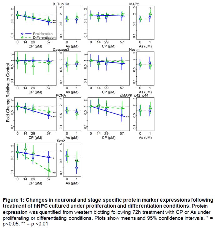
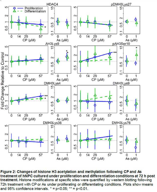
In conjunction with Dr. Costa, Dr. Faustman’s lab has also been examining developmental neurotoxic effects of OPs. In vitro models of neuronal differentiation are emerging as an important tool for high throughput and high content screening in neurodevelopmental toxicology. Understanding when, how, and at what doses neurotoxicants exposures affect normal development is critical for our ability to predict impacts of exposures before population-wide exposures occur. To further explore the importance of developmental context and timing in neurotoxicity we exposed human neuronal progenitor cells (hNPCs) grown in proliferating and differentiating conditions to chlorpyrifos (CP) and arsenic (As), two well established neurotoxicants. The effects of CP or As treatment on hNPC morphology and cell viability were measured 24 h and 72 h post-treatment and at 72 h post-treatment, changes in protein expression levels of neural differentiation and cell stress markers, cell viability and histone H3 modifications were observed (Figures 1 and 2). Cell viability, differentiation status, and epigenetic results suggest that hNPC cultures respond to CP and As treatment with different degrees of sensitivity, dependent on differentiation/proliferation status and on the toxicant concentration and length of exposure. Toxicant-related responses in sensitivity and protein expression patterns of neuronal markers that occurred 72 h post-treatment were dependent on the cell growth conditions. Histone modifications, as measured by changes in histone H3 phosphorylation, acetylation and methylation, varied for each toxicant and growth condition, suggesting that differentiation status can influence the epigenetic effects of CP and As exposures (Figure 2).
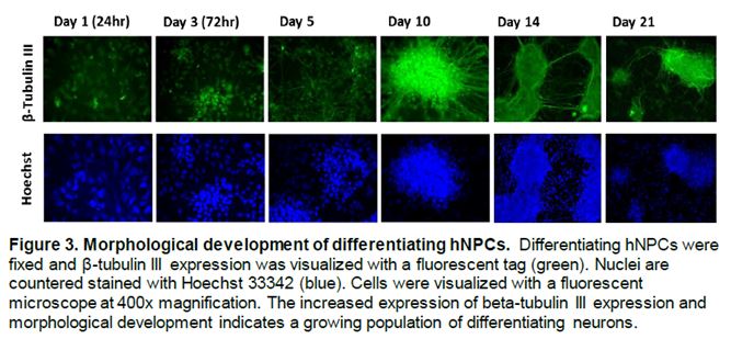
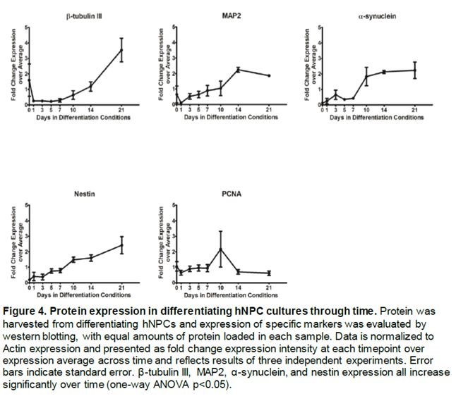
In addition, Dr. Faustman’s lab has characterized pathway dynamics throughout neuronal differentiation of the hNPC line that provides a particularly promising, scalable and reproducible model for high throughput and high content neurodevelopmental toxicity screening. The lab cultured hNPCs up to 21 days in differentiation conditions and used Western blotting and immunofluorescence to measure changes in protein expression though time (Figure 3 and 4). Global gene expression dynamics were measured using Affymetrix Human gene 2.0 ST microarrays. Over time in differentiation conditions, hNPCs acquired morphological characteristics of mature neuronal networks and increased expression of neuronal differentiation markers, including beta tubulin III, MAP2, and synaptophysin. Significantly changed genes were organized according to temporal expression patterns using K-means clustering, revealing 3 phases of gene expression. Quantitative pathway analysis identified gene ontology (GO) terms enriched among genes expressed in each of these phases and created a quantitative summary or temporal pathway trends in vitro. GO terms enriched among genes significantly decreased over time are largely associated with proliferation, and stem cell maintenance. GO terms enriched among genes with significantly increasing expression over time are dominated by key developmental processes, including neuronal differentiation, migration, and synaptogenesis. Enrichment of several GO terms associated with forebrain development indicates that these culture conditions promote differentiation towards a forebrain identity.
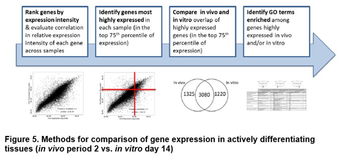
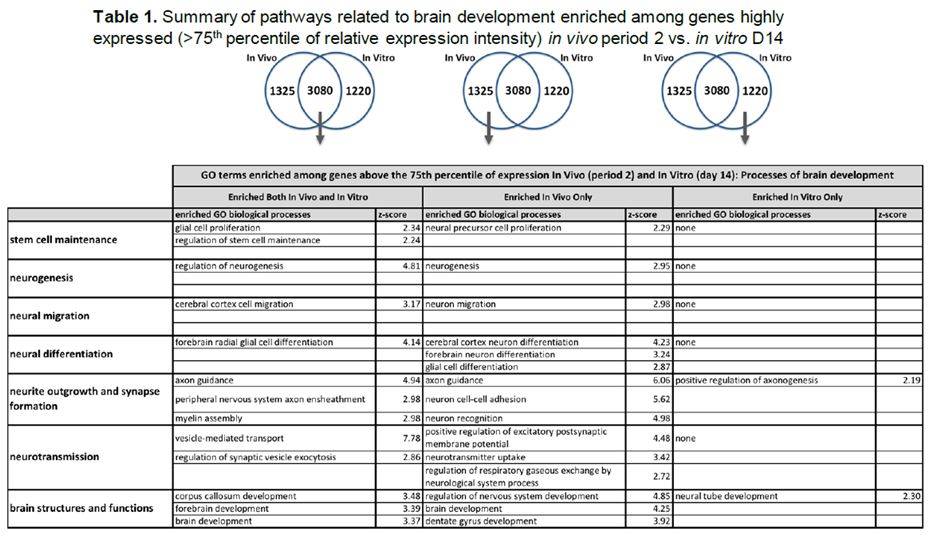
We compared gene expression in vitro with publicly available gene expression data from developing human brain tissue in vivo and found substantial concordance in relative gene expression intensity (Figure 5). Genes highly expressed in both samples were enriched for key processes of brain development, including proliferation, migration, differentiation, synapse formation, and neurotransmission (Table 1). Conversely, GO terms enriched among genes highly expressed only in vivo or only in vitro reveal important differences between systems. For example, genes highly expressed in vitro are enriched for more stress and apoptosis pathways. This analysis provides a timeline of progression through differentiation, facilitating identification of key phases of sensitivity in vitro. Key processes important for the identification of Adverse Outcome Pathways (AOPs) of proliferation, differentiation, and functional maturation matched in vivo patterns (Table 1). Given the heightened sensitivity of the brain to toxicant perturbation during critical windows of development, it is important that we understand which sensitive developmental pathways are captured in vitro and which are not so that in vitro assays can be interpreted appropriately. These observations of morphology, protein and gene expression provide a timeline of progression through differentiation, facilitating identification of key phases of sensitivity. By anchoring in vitro dynamics to in vivo reference points, this work clarifies the extent to which fundamental processes of brain development are captured in our model.
Significance
The focus of the Faustman and Costa laboratories is to understand the potential for, and magnitude of, impacts of pesticides on neurogenesis and gliogenesis. In both these essential neurodevelopmental pathways the balance between initial proliferation and subsequent specific differentiation is integral for proper neurodevelopment. In this project critical molecular pathways facilitating proliferation and toxicant response are being investigated. Knowledge about the timing and sensitivity of these critical pathways will directly translate into information relevant for establishing conditions promoting environmental and public health safety.
By anchoring in vitro gene expression dynamics to in vivo dynamics, the analysis above begins to define the appropriate applications of Dr. Costa’s in vitro model of neuronal differentiation for developmental neurotoxicology. The neuronal differentiation model evaluated here captures several essential processes of early brain development in vivo, including neuronal differentiation and development, neuronal migration, synapse formation, and neurotransmission. This model also captures several generic developmental pathways important throughout development. For example, pathway analysis revealed activity in signal transduction pathways and general differentiation and morphogenesis processes that are ubiquitous in development. Detection of perturbation of generic developmental pathways in this model may be able to predict perturbation in a broader set of developmental contexts.
Taken together, Dr. Faustman’s results support the idea that dose, time, and biological context are important factors that need to be considered when developing in vitro models for toxicity testing, interpreting results, and comparing findings across endpoints and platforms.
Key Center Findings Related to the Molecular Mechanisms Project
- Both diazinon and diazinon-oxon adversely affect astrocyte function, resulting in inhibited neurite outgrowth in hippocampal neurons. Inhibited outgrowth is associated with neurodegenerative diseases.
- Diazinon and diazinon-oxon increase oxidative stress in astrocytes, and this in turn modulates astrocytic fibronectin, leading to impaired neurite outgrowth in hippocampal neurons.
- Diazinon and diazinon-oxon significantly inhibited neurite outgrowth in hippocampal neurons, at concentrations devoid of cytotoxicity. These effects appeared to be mediated by oxidative stress, as they were prevented by antioxidants (melatonin, N-tbutyl- alpha-phenylnitrone, and glutathione ethyl ester).
- Inhibition of neurite outgrowth was observed at concentrations below those required to inhibit the catalytic activity of acetylcholinesterase. Diazinon and diazinon-oxon inhibit neurite outgrowth in hippocampal neurons by mechanisms involving oxidative stress, and that these effects can be modulated by astrocytes and astrocyte-derived GSH. These responses are similar to those observed following OP exposure.
- Differentiation status (or developmental context) modifies the epigenetic effects of chlorpyrifos and arsenic exposure.
- Characterization of neurodevelopmental toxicants using human neuroprogenitor cells provides a particularly promising, scalable and reproducible model for high throughput and high content neurodevelopmental toxicity screening.
We have identified common mechanisms of actions for OP Pesticides that have informed our analysis of adverse outcome pathways which link OPs to adverse neurodevelopmental outcomes.
Future Activities:
The Faustman and Costa labs will continue with their investigations to support the aims of the Molecular Mechanisms project and publish drafted manuscripts.
References:
Guizzetti M, Pathak S, Giordano D, Costa L. 2005. Effect of organophosporus insecticides and their metabolites on astroglial cell proliferation. Toxicology. Vol. 215:182-190.
Giordano G, Afsharinejad Z, Guizzetti M, Vitalone A, Kavanagh T, Costa L. 2007. Organophosphorus insecticides chlorpyrifos and diazinon and oxidative stress in neuronal cells in a genetic model of glutathione deficiency. Toxicology and Applied Pharmacology. Vol. 219:181-189
Guizzetti M, Moore NH, Giordano G, Costa LG. 2008. Modulation of neuritogenesis by astrocyte muscarinic receptors. J. Biol Chem. Vol. 283:1884-97.
Guizzetti M, More NH, Giordano G, VanDeMark KL, Costa LG. 2010. Ethanol inhibits neuritogenesis induced by astrocyte muscarinic receptors. Glia. Vol. 58:1395-1406.
Giordano G, Pizzuro D, VanDeMark K, Guizzetto M, Costa LG. 2009. Manganeze Inhibits the ability of astrocytes to promote neuronal differentiation. Toxicology and Applied Pharmacology. Vol. 240:226-235.
Pizzurro, D.M., K. Dao, and L.G. Costa, 2014a. Diazinon and diazoxon impair the ability of astrocytes to foster neurite outgrowth in primary hippocampal neurons. Toxicol Appl Pharmacol. 274(3): p. 372-82.
Pizzurro, D.M., K. Dao, and L.G. Costa, 2014b. Astrocytes protect against diazinon- and diazoxon-induced inhibition of neurite outgrowth by regulating neuronal glutathione. Toxicology. Vol. 318:59-68.
Journal Articles on this Report : 6 Displayed | Download in RIS Format
| Other subproject views: | All 55 publications | 38 publications in selected types | All 16 journal articles |
|---|---|---|---|
| Other center views: | All 510 publications | 227 publications in selected types | All 178 journal articles |
| Type | Citation | ||
|---|---|---|---|
|
|
Costa LG, Pellacani C, Dao K, Kavanagh TJ, Roque PJ. The brominated flame retardant BDE-47 causes oxidative stress and apoptotic cell death in vitro and in vivo in mice. NeuroToxicology 2015;48:68-76. |
R834514 (2015) R834514 (Final) R834514C003 (2015) R834514C003 (Final) |
Exit Exit Exit |
|
|
Harris S, Hermsen SA, Yu X, Hong SW, Faustman EM. Comparison of toxicogenomic responses to phthalate ester exposure in an organotypic testis co-culture model and responses observed in vivo. Reproductive Toxicology 2015;58:149-159. |
R834514 (Final) R834514C003 (2015) R834514C003 (Final) R835738 (2016) R835738 (2017) R835738C004 (2015) R835738C004 (2017) |
Exit Exit Exit |
|
|
Kim HY, Wegner SH, Van Ness KP, Park JJ, Pacheco SE, Workman T, Hong S, Griffith W, Faustman EM. Differential epigenetic effects of chlorpyrifos and arsenic in proliferating and differentiating human neural progenitor cells. Reproductive Toxicology 2016;65:212-223. |
R834514 (Final) R834514C003 (2015) R834514C003 (Final) |
Exit Exit Exit |
|
|
Wegner SH, Yu X, Pacheco Shubin S, Griffith WC, Faustman EM. Stage-specific signaling pathways during murine testis development and spermatogenesis: a pathway-based analysis to quantify developmental dynamics. Reproductive Toxicology 2015;51:31-39. |
R834514 (2015) R834514 (Final) R834514C003 (2015) R834514C003 (Final) R835738 (2016) R835738 (2017) R835738C004 (2015) R835738C004 (2017) |
Exit Exit Exit |
|
|
Wegner S, Yu X, Kim HY, Harris S, Griffith WC, Hong S, Faustman EM. Effect of dipentyl phthalate in 3-dimensional in vitro testis co-culture is attenuated by cyclooxygenase-2 inhibition. Journal of Toxicology and Environmental Health Sciences 2014;6(8):161-169. |
R834514 (2015) R834514 (Final) R834514C003 (2015) R834514C003 (Final) |
Exit Exit |
|
|
Zhou C, Chen J, Zhang X, Costa LG, Guizzetti M. Prenatal ethanol exposure up-regulates the cholesterol transporters ATP-binding cassette A1 and G1 and reduces cholesterol levels in the developing rat brain. Alcohol and Alcoholism 2014;49(6):626-634. |
R834514 (2015) R834514 (Final) R834514C003 (2015) R834514C003 (Final) |
Exit Exit Exit |
Supplemental Keywords:
Health, RFA, Scientific Discipline, INTERNATIONAL COOPERATION, ENVIRONMENTAL MANAGEMENT, Environmental Policy, Biology, Children's Health, Biochemistry, Environmental Monitoring, Risk Assessment, biological markers, pesticides, age-related differences, pesticide exposure, agricultural community, children's vulnerablity, molecular researchProgress and Final Reports:
Original AbstractMain Center Abstract and Reports:
R834514 Predictive Toxicology Center for Organotypic Cultures and Assessment of AOPs for Engineered Nanomaterials Subprojects under this Center: (EPA does not fund or establish subprojects; EPA awards and manages the overall grant for this center).
R834514C001 Community-Based Participatory Research
R834514C002 Pesticide Exposure Pathways
R834514C003 Molecular Mechanisms
R834514C004 Genetic Susceptibility
The perspectives, information and conclusions conveyed in research project abstracts, progress reports, final reports, journal abstracts and journal publications convey the viewpoints of the principal investigator and may not represent the views and policies of ORD and EPA. Conclusions drawn by the principal investigators have not been reviewed by the Agency.