
Grantee Research Project Results
2010 Progress Report: Project 5 -- Architecture Development and Particle Deposition
EPA Grant Number: R832414C005Subproject: this is subproject number 005 , established and managed by the Center Director under grant R832414
(EPA does not fund or establish subprojects; EPA awards and manages the overall grant for this center).
Center: Center for the Study of Metals in the Environment
Center Director: Allen, Herbert E.
Title: Project 5 -- Architecture Development and Particle Deposition
Investigators: Wexler, Anthony S. , Winkle, Laura Van , Plopper, Charles , Schelegle, Ed
Current Investigators: Wexler, Anthony S. , Plopper, Charles
Institution: University of California - Davis
EPA Project Officer: Chung, Serena
Project Period: October 1, 2005 through September 30, 2010 (Extended to September 30, 2011)
Project Period Covered by this Report: July 1, 2009 through June 30,2010
RFA: Particulate Matter Research Centers (2004) RFA Text | Recipients Lists
Research Category: Human Health , Air
Objective:
Quantify lung architecture, pulmonary function and particle deposition pattern changes due to pollutant exposure during development.
Progress Summary:
Three current activities are highlighted below: (1) lung architecture and function changes due to premixed flame particle with NMAD 212 nm (PFP212) exposure during development, (2) cell proliferation related gene and protein expression examination after a single exposure to PFP73, PFP212 and DPF230 at 7 days of age, and (3) lung architecture and function changes due to ozone (2 days exposure and 5 days recovery) plus premixed flame particle (PFP73) exposure during development.
1. Lung architecture, function changes due to PFP212 exposure and cell proliferation gene expression change due to DFP230, PFP73, PFP212 during development
The diffusion flame produced a concentration of 2.4x10⁴ particles/cm3 in the exposure chamber with a number mean aerodynamic particle diameter (NMAD) of 230 nm and a mass concentration of 71.7 µg/m3 (DFP230). The premixed flames with φ=2.2, 2.5 produced 9.5x10⁴, 4.3x10⁴ particles/cm3 in the chamber with a diameter (NMAD) of 72.7, 212.0 nm and a mass concentration of 20.0, 67.4 µg/m3, respectively, which we refer to as PFP73 and PFP212. Litters of male Sprague-Dawley rat pups housed with lactating mothers were placed in filtered air chambers at age one day. Exposure for six hours a day, five days a week for three weeks began when the pups were seven days old and ended at 25 days old. A matched set of pups was exposed to filtered air using the same protocol. At 28 days old, the animals were transferred from the exposure chambers to Bioclean hoods where they matured until necropsy at 80-81 days of age.
Total lung capacity (TLC) normalized by body weights (BW) and BW itself were similar between FA group and exposed groups (p=0.8, 0.277, 0.076 for DFP230, PFP73 and PFP212 respectively) at age of 80 or 81 days.
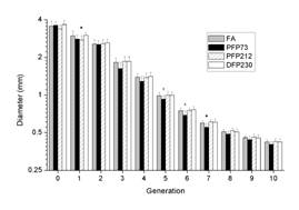
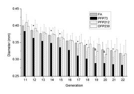
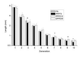
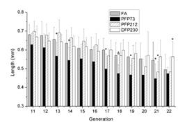
Figures 1 and 2 show the generation-averaged airway diameter and length. Contrary to PFP73, there were no significant differences in airway diameter and length between the FA and PFP212.
 |
 |
| (a) | (b) |
Figure 1. Airway diameter as a function of generation number in the different groups (a) from generation 0 to 10, (b) from generation 11 to 22. * indicates p-value less than 0.05 and & indicates p-value less than 0.0022 (Bonferroni adjustment).
 |
 |
| (a) | (b) |
Figure 2. Airway length as a function of generation number in the different groups (a) from generation 0 to 10, (b) from generation 11 to 22.
Exposure to PFP212 resulted in a significant increase (p = 0.013) in the methacholine EC200R indicating decrease in airway responsiveness in this group. Exposure to DFP230 or PFP73 did not significantly alter airway responsiveness to methacholine challenge when compared to FA. Exposure to PFP212 resulted in a significant increase in Rs (p = 0.005) and I (p = 0.001) compared to FA exposure. In contrast, exposure resulted in a significant decrease in I in the PFP73 (p = 0.001) group compared to FA. In addition, exposure to DFP230 resulted in a significant increase in H compared to FA exposure (p = 0.002).
Proliferating Cell Nuclear Antigen (PCNA), a gene associated with cell proliferation was quantitatively assayed using RT-PCR. Gene expression significantly decreased in neonatal rat airways following a single 6-hour exposure to PFP73 (p=0.0034), increased subsequent to PFP212 (p<0.0001) and remained unchanged from DFP230 (p=0.0675), as compared with FA control animals (Figure 3A). To qualitatively measure protein abundance, paraformaldehyde fixed lung sections were stained against PCNA immunohistochemically. Basal PCNA positive cells, identified as cells containing dense nuclear staining, were abundant across both airway epithelium and the peribronchiolar interstitium (Figure 3B). Abundance was diminished in PFP212 (Figure 3C) and DFP230 (Figure 3E) groups, but a plethora of PCNA positive proliferating cells were observed in the PFP73 group (Figure 3D).
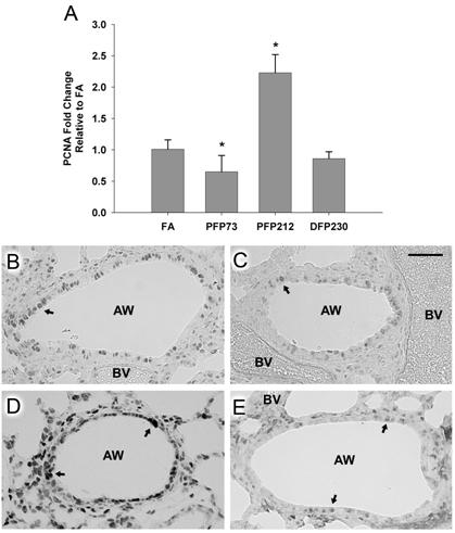
Figure 3. Expression of Proliferating Cell Nuclear Antigen (PCNA) mRNA and protein in the airways of neonatal 7 day old rats were evaluated. Gene expression was measured in microdissected airways using qRT-PCR and results were calculated using the comparative Ct method. GAPD was the reference gene. * denotes significance at P<0.05.
2. Lung architecture and function changes due to periodic exposure to Ozone plus PFP73 exposure during development (52/25)
This experiment was designed to investigate the effects of periodic exposure to ozone plus PFP73. Male Sprague Dawley rats were exposed to 0.5 ppm ozone plus PFP73 from 7 days to 26 days postnatal, 6 hrs/day, 2 days exposure and 5 days off for ozone and 5 days exposure and 2 days off for PFP73.
No significant geometric changes were elicited by co-exposure of ozone with PFP73. Exposure to 52/25 resulted in a significant decrease in the methacholine EC200R indicating increase in airway responsiveness in this group.
3. Measuring particle deposition in rat airways
Recently, we developed methods for identifying airway geometry in the conducting airways. The method is described by Lee and coworkers (2008a) and used by Lee and coworkers (2008b) to identify geometry and variability in 6 adult Sprague-Dawley rats. In this method, the rat lungs are cast using a mixture of silicone and oil, where the lung is bleached once the silicone has cured. A micro-CT scanner is used to scan the cast lung, and in house software is used to generate detailed airway characteristics. The availability of methods and software for quantifying the airway tree, led us to explore the possibility of imaging the airways and the particles deposited in them so that we could quantify airway-by-airway particle deposition.
One of the major shortcomings of using a micro-CT scanner to image deposited particles is that we cannot ensure deposited particles to stay in place during the process of casting, bleaching and imaging. A block-face imaging cryomicrotome from Barlow Scientific, Inc. can be used to image the airways and particles (Barlow and Kelly, 1997; Kelly et al., 2000). The imaging cryomicrotome serially sections through frozen organs and images the exposed surface using various excitation and emission filters. Slice thickness and imaging resolution can be varied using software and hardware controls to image up to four different fluorescent colors. The imaging method produces voxels as small as 10 um so tissue boundaries and particle locations could conceivably by imaged to this accuracy. With respect to the initial casting process, the process of casting organs for imaging with a cryomicrotome has been modified to ensure that particles are immobile.
Method Validation
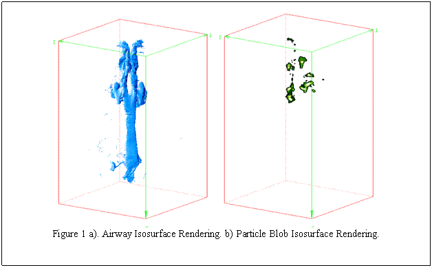
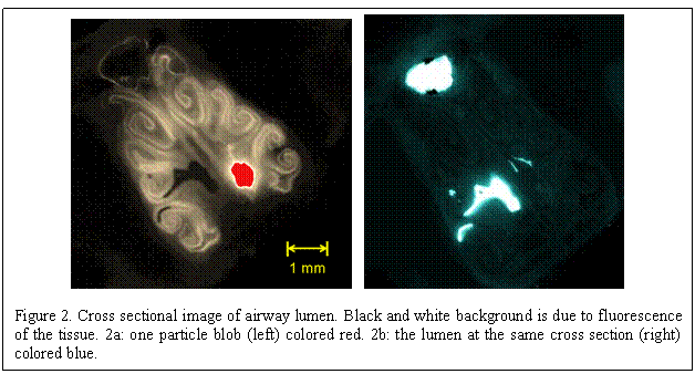
Rat nasal airways are quite complex but much less so than the conducting airways and the nasal airway length scales are much larger than the conducting airways so they are an easier place to begin this work. To find the detailed location of the particles in the nasal airways the surrounding tissue should not be bleached and the particles should not move during the scanning process. Over the course of many experiments we have come up with a technique of casting the nasal airways to freeze the deposited particles in place. In addition, it is easier to validate the method in the nasal airways because they are much more accessible. To validate the method, we used a hypodermic needle to place fluorescent particles at a few locations in the nasal passage of a rat airway. We then used our procedure to image the nasal passage and the particle location to ensure that the imaging technique located the particles in the same location where we placed them.
Figure 1a shows this rat’s reconstructed nasal airways viewed from two different viewpoints. Figure 1b shows the reconstruction of the particle blobs injected into the nasal cavity, viewed from the same vantage points as Figure 1a. Figure 2a shows a blowup of one of the particle blobs with tissue fluorescence in the background. Figure 2b shows an image of the lumen in the same plane as the blob. Our experiments with injecting the yellow spots in the rat nose and casting the airway with a silicone mixture shows that the method is feasible to detect particles in the airways.
Imaging rat nasal airways with deposited particles
After the new method for ensuring deposited particles are immobile and stationery during the entire process of necropsy, casting and slicing, an adult rat was exposed to aerosolized Dayglo yellow dye particles for 2 hrs using a aerosol dust generator (Carvacho 2001). The rat was placed in a nose only exposure chamber (Teague 2005). Figure 3 shows the exposure setup, Saturn yellow dye is placed inside the aerosol generator which is aerosolized, the particles pass through an electrostatic neutralizer to remove any charge buildup. The rat breathes the particles flowing into the chamber which is constantly being supplied with air. Rats were exposed to aerosolized yellow dye particles for a period of 2 hours with particle concentration of 1 mg/m3 (approx) monitored using a TSI Dusttrak. The Rat head is cast using a silicone mixture and allowed to cure, once cured the specimen is then shipped frozen to Barlow Scientific for imaging.
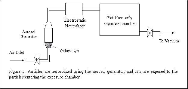
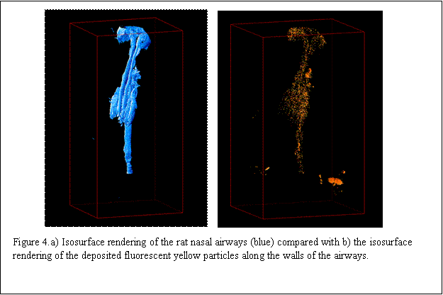
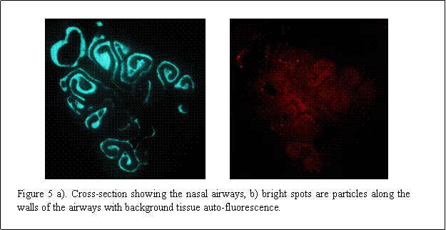
Figure 4a shows the rendering of the rat nasal airway and Figure 4b the deposited yellow particles along the airway. In Figure 5a, a cross-section of the airway is shown, and Figure 5b shows the deposited yellow particles as bright spots with background auto-fluorescence. These images clearly indicate that we can image deposited particles in the nasal airways.
Future Activities:
Experiments will be undertaken using particles of various sizes and colors, to determine where the particles are depositing in the nasal airways, and also the clearance of particles over time will be studied. PFP73 exposures with different concentration and exposure to maternal separation (social stress) have been completed and data analyses are currently underway.
References:
Journal Articles on this Report : 1 Displayed | Download in RIS Format
| Other subproject views: | All 21 publications | 13 publications in selected types | All 13 journal articles |
|---|---|---|---|
| Other center views: | All 128 publications | 71 publications in selected types | All 64 journal articles |
| Type | Citation | ||
|---|---|---|---|
|
|
Lee DY, Wallis C, Wexler AS, Schelegle ES, Van Winkle LS, Plopper CG, Fanucchi MV, Kumfer B, Kennedy IM, Chan JKW. Small particles disrupt postnatal airway development. Journal of Applied Physiology 2010;109(4):1115-1124. |
R832414 (Final) R832414C001 (2010) R832414C005 (2010) R832414C005 (Final) |
Exit Exit Exit |
Supplemental Keywords:
Health, RFA, Air, Scientific Discipline, PHYSICAL ASPECTS, ENVIRONMENTAL MANAGEMENT, Health Risk Assessment, Physical Processes, Risk Assessments, particulate matter, Environmental Chemistry, Biochemistry, Risk Assessment, particulate matter components, biological mechanisms, chemical characteristics, airborne particulate matter, human exposure, cardiopulmonary responses, animal model, children's health, exposure, PM, atmospheric particulate matter, toxicology, acute cardiovascular effects, human health effects, cardiovascular disease, human health risk, exposure assessmentProgress and Final Reports:
Original AbstractMain Center Abstract and Reports:
R832414 Center for the Study of Metals in the Environment Subprojects under this Center: (EPA does not fund or establish subprojects; EPA awards and manages the overall grant for this center).
R832414C001 Project 1 -- Pulmonary Metabolic Response
R832414C002 Endothelial Cell Responses to PM—In Vitro and In Vivo
R832414C003 Project 3 -- Inhalation Exposure Assessment of San Joaquin Valley Aerosol
R832414C004 Project 4 -- Transport and Fate Particles
R832414C005 Project 5 -- Architecture Development and Particle Deposition
The perspectives, information and conclusions conveyed in research project abstracts, progress reports, final reports, journal abstracts and journal publications convey the viewpoints of the principal investigator and may not represent the views and policies of ORD and EPA. Conclusions drawn by the principal investigators have not been reviewed by the Agency.