U.S. flag

An official website of the United States government

Here’s how you know

Dot gov

Official websites use .gov
A .gov website belongs to an official government organization in the United States.

HTTPS

Secure .gov websites use HTTPS
A lock (LockA locked padlock) or https:// means you have safely connected to the .gov website. Share sensitive information only on official, secure websites.

  • Environmental Topics
  • Laws & Regulations
  • Report a Violation
  • About EPA
Contact Us

Grantee Research Project Results

2012 Progress Report: Multi-Sensor Reporter Cell Technology to Assess Hazard Involving Endocrine Signaling Pathways

EPA Grant Number: R835165
Title: Multi-Sensor Reporter Cell Technology to Assess Hazard Involving Endocrine Signaling Pathways
Investigators: LeBlanc, Gerald A.
Institution: North Carolina State University
EPA Project Officer: Aja, Hayley
Project Period: March 1, 2012 through February 28, 2016 (Extended to February 28, 2017)
Project Period Covered by this Report: March 1, 2012 through February 28,2013
Project Amount: $950,507
RFA: Developing High-Throughput Assays for Predictive Modeling of Reproductive and Developmental Toxicity Modulated Through the Endocrine System or Pertinent Pathways in Humans and Species Relevant to Ecological Risk Assessment (2011) RFA Text |  Recipients Lists
Research Category: Chemical Safety for Sustainability

Objective:

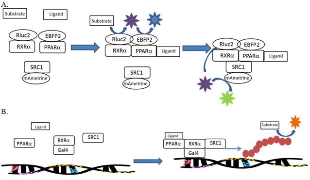
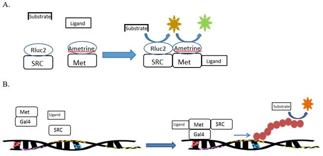
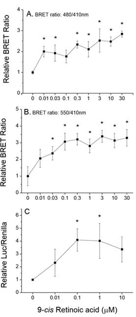
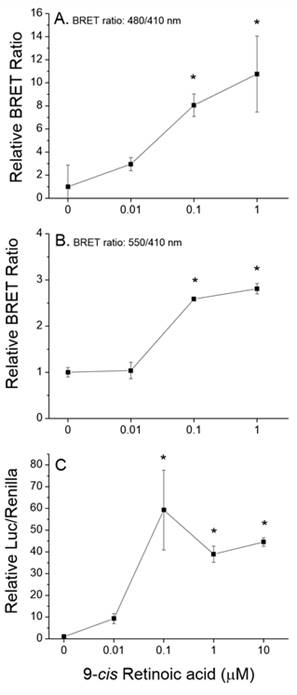
The high-throughput evaluation of toxicity pathways is an emerging paradigm for future toxicity characterization. In order to meet the goals of this paradigm, methods are needed to assess toxicity pathways using as few assays as possible. We propose the use of multi-sensor cell-based reporter assays to meet this need. These assays will utilize both bioluminescence resonance energy transfer (BRET) and reporter gene assays to evaluate interactions of individual chemicals and chemical mixtures along nuclear receptor-mediated signaling pathways. Methods are being developed for the evaluation of chemical effects on the PPARα:RXRα:SRC1 signaling pathway, PPARγ:RXRα:SRC1 signaling pathway, RXRα:RXRα:SRC1 signaling pathway, and the MET:SRC signaling pathway. The former three pathways utilize human receptors that regulate various aspects of glucose and lipid homeostasis. The latter pathway utilizes receptors from the invertebrate Daphnia that regulates various aspects of sex determination and reproductive maturation in arthropods. The development of a high-throughput assay that would identify specific toxicological targets within signaling networks would provide a wealth of mechanistic information on the potential hazard of individual chemicals. This mechanistic data could then be used to model toxicity associated with chemical mixtures.

Progress Summary:

Aim 1. Construct a next-generation, multi-sensor reporter assay for the detection of chemical interactions with the vertebrate RXR:PPAR signaling pathway. Effort during year 1 was directed primarily towards the construction of the assay components and development of the assay procedures. The overall objectives of this program were expanded over that originally proposed, as the initial proposal was to develop the screening methods using a single signaling pathway: PPARα:RXRα:SRC1. The program was expanded to include three additional pathways: PPARγ:RXRα:SRC1, RXRα:RXRα:SRC1, and MET:SRC.
 
Construction of the human PPAR, RXR, and SRC1 fusion proteins
 
The general premise behind the assay design was that BRET signals would be used to assess dimerization between receptor subunits with commensurate recruitment of the coactivator SRC1 following addition of the test chemicals. Two hybrid assays would be used to assess activation of the overall complex in response to added test chemical.
 
The following fusion proteins were created for assessment in the BRET and reporter gene assays:
  1. gal4-RXRα(DEF)-Rluc2
  2. PPARα(ORF)-EBFP2
  3. gal4-PPARα(DEF)
  4. RXRα(ORF)-EBFP2
  5. SRC1(ORF)-mAmetrine
  6. PPARγ(ORF)-EBFP2
  7. gal4-PPARγ(DEF)
The gal4 DNA binding sites were fused to the proteins to provide for a DNA-binding subunit during two hybrid assays. Renillaluciferase 2 (Rluc2) was fused to RXRα to serve as the photon donor (410 nm) during BRET assays. Enhanced blue fluorescent protein 2 (EBFP2) was fused to proteins that would partner with RXRα. EBFP2 would serve as the fluorescent photon acceptor during BRET assays (480 nm emission). mAmetrine was fused to SRC1 to serve as the fluorescent photon acceptor during BRET assays (550 nm emission). Fluorescent proteins were fused to both the c-terminus and the n-terminus of the proteins. Constructs that provided the strongest BRET signal were then selected for routine use.
 
Assay development
 
Assays involving the human receptor proteins were developed using HepG2 cells as the assay host. Efforts to develop a single assay that incorporated both BRET and reporter gene endpoints were unsuccessful due to incompatible assay requirements. Therefore, methods were developed for either the concurrent or sequential use of the BRET and reporter gene assays. Methods were initially developed using the ligand tributyltin (TBT), as TBT is a ligand to RXRα, PPARα and PPARγ. BRET and reporter-gene assay designs are illustrated in Fig. 1.
 
Construction of the daphnid MET and SRC fusion proteins
 
The general premise behind theisassay design was that a BRET signal would be used to assess dimerization of MET and SRC following addition of the test chemical. The two hybrid assays would be used to assess activation of the receptor complex in response to added test chemical.
 
The assay involving the daphnid proteins were developed using Drosophila S2 cells as the assay host. The daphnid Met gene was previously cloned in our lab, however, the daphnid SRC required cloning. Cloning of this formidable gene (7,074 base pairs) was accomplished during the first year of this program. The following fusion proteins were subsequently generated:
  1. mAmetrine-MET-GAL4
  2. GAL4-MET-mAmetrine
  3. Rluc2-SRC
  4. SRC-SRC-Rluc2
All fusion protein constructs (c-terminus and n-terminus fusions) were evaluated to identify the combination that provided the strongest BRET signal. This combination was then used in all BRET assays. BRET and reporter gene designs are illustrated in Fig. 2.
 
Figure 1. A. Illustration of the BRET assay designed to detect dimerization of RXRα and PPARα along with recruitment of SRC1. B. Reporter gene assay designed to measure the ability of the activated receptor complex to stimulate gene transcription.
 
Figure 1
 
 
Figure 2. A. Illustration of the BRET assay designed to detect dimerization of MET and SRC. B. Reporter gene assay designed to measure the ability of the activated receptor complex to stimulate gene transcription.
 
Figure 2
 
Aim 2. Functionally validate the assay using multiple chemicals that impact different targets along the RXR:PPAR signaling pathway. Assays involving the PPARα:RXRα and RXRα:RXRα signaling pathways were evaluated using the well characterized RXRα ligands 9-cis retinoic acid and LGD1069.
 
RXRα ligands BRET experiments were performed, with RXRα fused to a photon donor protein (Rluc2) and PPARα fused to a fluorescent protein (EBFP2), to evaluate liganddependent dimerization of the nuclear receptor subunits. Subunit dimerization increased with increasing 9-cis retinoic acid concentration (Fig. 3A). Commensurate with this dimerization was the recruitment of co-activator SRC1, which was fused to mAmetine fluorescent protein (Fig. 3B). Thus, binding of 9-cis retinoic acid to RXRα simulated the formation of this nuclear receptor triplex. This triplex functioned as a competent transcription factor, as the reporter gene was activated in the reporter gene assay by this complex in a 9-cis retinoic acid-concentration dependent manner (Fig. 3C). Similar experiments that assessed RXRα:PPARα:SRC1 complexation and activation in response to the alternative RXRα ligand LGD1069 yielded similar results.
 
The ability of 9-cis retinoic acid to stimulate dimerization of two RXRα subunits along with SRC1 recruitment to the homodimer also was evaluated. Here, one-half of RXRα protein expressed in the assay was fused to the photon donor Rluc2, and the other half was fused to the fluorescent protein EBFP2. Again, subunit dimerization (Fig. 4A), co-activator recruitment (Fig. 4B), and receptor activation (Fig. 4C) all were stimulated in a 9-cis retinoic acid concentration-dependent manner. Taken together, results demonstrate that ligand occupancy of human RXRα stimulates heterodimerization with PPARα and homodimerization along with recruitment of SRC1 to the dimers. Further, these dimerization evaluations were performed in the absence of a DNA docking site indicating that dimerization and recruitment occur independent of association with DNA.
 
PPARα ligands Receptor subunit heterodimerization and SRC1 recruitment to the dimer in response to the PPARα ligand clofibrate was evaluated using BRET. Clofibrate did not stimulate subunit heterodimerization (Fig. 5A) or SRC1 recruitment (Fig. 5B). Yet, clofibrate did stimulate transcriptional activation by the complex (Fig. 5C). Similar experiments performed with an alternative PPARα ligand, Wy14643, also failed to stimulate dimerization of RXRα and PPARα and recruitment of SRC1 but did stimulate transcriptional activation by the complex. Transcriptional activation by the subunit heterodimer, and not a PPARα monomer or homodimer, is evidenced by the fact that DNA binding was a characteristic of the RXRα subunit (which contained the gal-4 DNA binding site); while, PPARα contained the ligand binding site. Thus, while RXRα ligands stimulate dimerization and co-activator recruitment, PPARα ligands activate constitutively existing subunit heterodimers. The activation of existing subunit heterodimers by PPARα ligands was further supported by the observation that basal, constitutive transcriptional activation was low in reporter assays containing only gal-4RXRα or gal-4-PPARα, but significantly increased when both proteins were expressed together (Fig. 6). As expected, clofibrate did not stimulate RXRα homodimer formation (Fig. 7A), recruitment of SRC1 (Fig. 7B), or transcriptional activation by the RXRα homodimer (Fig. 7C).
 
 
Figure 3
Figure 3. 9-cis retinoic acid-mediated (A) dimerization of RXRα and PPARα, (B) recruitment of SRC1 to the complex, and (C) transcriptional activation of a luciferase reporter gene by the complex. Data are presented as mean and standard deviation (n=3) normalized to control values. An asterisk denotes a significant (p<0.05) difference from the control (ANOVA, Tukey’s Post Hoc Test).
 
 
Figure 4
Figure 4. 9-cis retinoic acid-mediated (A) homodimerization of RXRα, (B) recruitment of SRC1 to the complex, and (C) transcriptional activation of a luciferase reporter gene by the complex. Data are presented as mean and standard deviation (n=3) normalized to control values. An asterisk denotes a significant (p<0.05) difference from the control (ANOVA, Tukey’s Post Hoc Test).
 
 
Figure 5
 
Figure 5. Effect of clofibrate on (A) dimerization of RXRα and PPARα, (B) Recruitment of SRC1 to the complex, and (C) transcriptional activation of a luciferase reporter gene by the complex. Data are presented as mean and standard deviation (n=3) normalized to control values. An asterisk denotes a significant (p<0.05) difference from the control (ANOVA, Tukey’s Post Hoc Test).
 
 
Figure 6
Figure 6. Ligand-independent activation of the luciferase reporter gene in the presence of gal4-RXRα, gal4-PPARα or both proteins together. Data are presented as mean and standard deviation (n=3). An asterisk denotes a significant difference from other treatments (ANOVA, Tukey’s Post Hoc Test).
 
 
Figure 7
Figure 7. Effect of clofibrate on (A) homodimerization of RXRα, (B) recruitment of SRC1 to the complex, and (C) transcriptional activation of a luciferase reporter gene by the complex. Data are presented as mean and standard deviation (n=3) normalized to control values.

Future Activities:

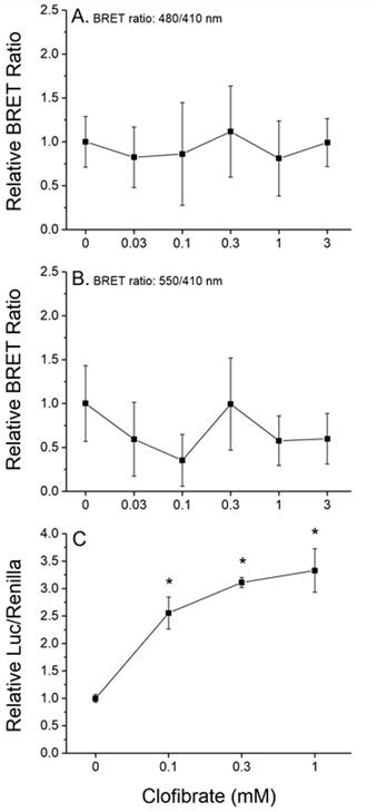
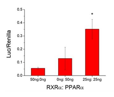
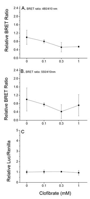
Assays were designed, constructed, and evaluated for the screening of chemicals for activity towards two additional signaling pathways: human PPARα:RXRα:SRC1 and daphnid MET-SRC. These assays will be validated during year 2 of the program. 

Journal Articles:

No journal articles submitted with this report: View all 26 publications for this project

Supplemental Keywords:

Multi-sensor reporter cell signaling, signaling pathways, endocrine disruptors, assays, ligands

Progress and Final Reports:

Original Abstract
  • 2013 Progress Report
  • 2014 Progress Report
  • 2015 Progress Report
  • Final Report
  • Top of Page

    The perspectives, information and conclusions conveyed in research project abstracts, progress reports, final reports, journal abstracts and journal publications convey the viewpoints of the principal investigator and may not represent the views and policies of ORD and EPA. Conclusions drawn by the principal investigators have not been reviewed by the Agency.

    Project Research Results

    • Final Report
    • 2015 Progress Report
    • 2014 Progress Report
    • 2013 Progress Report
    • Original Abstract
    26 publications for this project
    7 journal articles for this project

    Site Navigation

    • Grantee Research Project Results Home
    • Grantee Research Project Results Basic Search
    • Grantee Research Project Results Advanced Search
    • Grantee Research Project Results Fielded Search
    • Publication search
    • EPA Regional Search

    Related Information

    • Search Help
    • About our data collection
    • Research Grants
    • P3: Student Design Competition
    • Research Fellowships
    • Small Business Innovation Research (SBIR)
    Contact Us to ask a question, provide feedback, or report a problem.
    Last updated April 28, 2023
    United States Environmental Protection Agency

    Discover.

    • Accessibility
    • Budget & Performance
    • Contracting
    • EPA www Web Snapshot
    • Grants
    • No FEAR Act Data
    • Plain Writing
    • Privacy
    • Privacy and Security Notice

    Connect.

    • Data.gov
    • Inspector General
    • Jobs
    • Newsroom
    • Open Government
    • Regulations.gov
    • Subscribe
    • USA.gov
    • White House

    Ask.

    • Contact EPA
    • EPA Disclaimers
    • Hotlines
    • FOIA Requests
    • Frequent Questions

    Follow.