
Grantee Research Project Results
2009 Progress Report: Assessment of Microbial Pathogens in Drinking Water using Molecular Methods Coupled with Solid Phase Cytometry
EPA Grant Number: R833830Title: Assessment of Microbial Pathogens in Drinking Water using Molecular Methods Coupled with Solid Phase Cytometry
Investigators: Pyle, Barry H , Ford, Timothy E.
Current Investigators: Pyle, Barry H , Camper, Anne
Institution: Montana State University - Bozeman , Little Big Horn College
EPA Project Officer: Aja, Hayley
Project Period: March 1, 2008 through February 28, 2011 (Extended to February 28, 2013)
Project Period Covered by this Report: June 10, 2009 through February 28,2010
Project Amount: $599,996
RFA: Development and Evaluation of Innovative Approaches for the Quantitative Assessment of Pathogens and Cyanobacteria and Their Toxins in Drinking Water (2007) RFA Text | Recipients Lists
Research Category: Drinking Water , Water
Objective:
To develop and evaluate innovative approaches for quantitative assessment of pathogens in drinking water sources.
Progress Summary:
- Escherichia coli O157:H7
- Helicobacter pylori
- Legionella pneumophila
- Mycobacterium avium
- Aeromonas hydrophila
- Giardia lamblia
- Cryptosporidium parvum
Procedures
- Fluorescent in situ hybridization (FISH): enhance with tyramide amplification (TSA), labels with increased fluorescent intensity and/or use polyamide nucleic acid (PNA) probes.
- In situ nucleic acid amplification: Specific target genes inside individual cells (Hodson et al, 1995); use improved methods, e.g. (Notomi et al, 2000; Maruyama et al, 2003 & 2005), with membrane filtration and Solid Phase Laser Cytometry (SPLC).
SPLC using ScanRDI (AES-Chemunex)
- Scan a 25 mm diameter membrane filter in 3-4 minutes
- Detect individual fluorescent particles
- Discriminate between cells and debris
- Locate particles on microscope
- Validate bacteria, eliminate other particles
Cell Labels used with the ScanRDI
- Total Cell Count - Sybr Green
- Total Viable Count – ChemChrome detects enzyme activity and membrane integrity
- Identification Tests – Antibodies, specific enzymes, nucleic acid probes, FISH
- Dual Labeling - Fab-CTC, ChemChrome-Fab, DVC-FISH (Baudart et al, 2002)
Initial Work
E. coli was used with EUB338, a universal eubacterial DNA probe labeled with either HRP for TSA amplification (Schonhuber et al 1997), Alexa dye, or fluorescein; ECO541, an E. coli specific probe labeled with Alexa; and Unibact1, a universal eubacterial PNA probe tagged with fluorescein.
Fluorescent in situ Hybridization (FISH)
Fixation methods Combinations of ethanol baths, para formaldehyde and lysozyme treatment have been examined. It is necessary to use proprietary AES Chemunex polyester filters (CB 04) for this procedure as they resist removal of the black dye by the fixation reagents.
Temperature Hybridization for 2 hours at 46°C was optimal for probing with both EUB-HRP and ECO-Alexa.
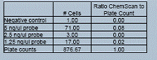
Table 1 Cells labeled with ECO-Alexa probe enumerated by ScanRDI compared to plate counts.
Enumeration with epifluorescent microscopy
Total cell counts with SYBR Green and FISH were done after enhanced labeling with chloramphenicol, TSA, and alternate probes.
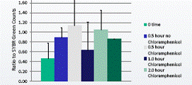
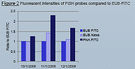
Figure 1 Ratio of cell numbers using FISH probe
ECO-Alexa 488 to SYBR Green after Chloramphenicol incubation to amplify RNA (Ouverney et al 1997).
Discussion
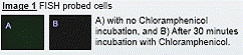
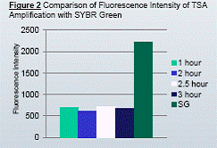
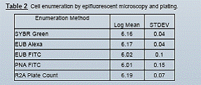
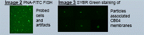
When E. coli cells were labeled with FISH probes tagged with Alexa dye and no additional enhancements, the ScanRDI enumerated up to about 8 percent of the cells present on a membrane (Table 1). Modified techniques have been tested to increase fluorescent intensity without introducing confounding particles. A short preincubation with chloramphenicol enhanced the fluorescent intensity of labeled probes by increasing the relative number of targeted ribosomes in the E. coli cells (Fig. 1 and Image 1). TSA also increased the fluorescence by enzymatically increasing the number of specific fluorescein molecules in the cells (Fig. 2). These two methods could be used in combination to produce cells that have sufficient fluorescence to be reliably enumerated by the ScanRDI. An initial comparison between DNA probes tagged with Alexa 488 or fluorescein (FITC) and PNA probes with FITC showed that while PNA probes, due to their uncharged backbone, enter the cells more reliably than similar DNA probes, the number of particulates in these preparations precluded the use of these probes for enumeration by the ScanRDI (Image 2). Alexa 488 is brighter than FITC without TSA, but not as intense as cells after the amplification. Enumeration by epifluorescent microscopy of SYBR Green stained cells and FISH labeled cells give similar results to plate counts, indicating that the FISH methods are reliably labeling all cells present (Table 2).
Problems have occurred when attempting to compare FISH probed cells with total cells stained by ScanRDI due to a large number of stained particles, between 1500 and 6000 particles per membrane (Image 3). Numerous procedures have been tried to eliminate these particles with little success. An alternative staining technique is being considered to overcome this challenge.
Future Activities:
References:
Journal Articles:
No journal articles submitted with this report: View all 6 publications for this projectProgress and Final Reports:
Original AbstractThe perspectives, information and conclusions conveyed in research project abstracts, progress reports, final reports, journal abstracts and journal publications convey the viewpoints of the principal investigator and may not represent the views and policies of ORD and EPA. Conclusions drawn by the principal investigators have not been reviewed by the Agency.