U.S. flag

An official website of the United States government

Here’s how you know

Dot gov

Official websites use .gov
A .gov website belongs to an official government organization in the United States.

HTTPS

Secure .gov websites use HTTPS
A lock (LockA locked padlock) or https:// means you have safely connected to the .gov website. Share sensitive information only on official, secure websites.

  • Environmental Topics
  • Laws & Regulations
  • Report a Violation
  • About EPA
Contact Us

Grantee Research Project Results

Final Report: Rapid Concentration, Detection, and Quantification of Pathogens in Drinking Water

EPA Grant Number: R833840
Title: Rapid Concentration, Detection, and Quantification of Pathogens in Drinking Water
Investigators: Hu, Zhiqiang , Riley, Lela K. , Lin, Mengshi
Institution: University of Missouri - Columbia
EPA Project Officer: Aja, Hayley
Project Period: May 1, 2008 through April 30, 2011 (Extended to April 30, 2013)
Project Amount: $600,000
RFA: Development and Evaluation of Innovative Approaches for the Quantitative Assessment of Pathogens and Cyanobacteria and Their Toxins in Drinking Water (2007) RFA Text |  Recipients Lists
Research Category: Drinking Water , Water

Objective:

Pathogens in drinking water and their associated waterborne disease are of great concern to the public health. Numerous molecular technologies and methods have been developed and are being applied to pathogen determination in drinking water. Most of these methods, however, require sample pretreatment to concentrate the bacteria or viruses in water before analysis. A common method to accomplish this requirement is filtration using a large volume of test water. The filtration process can be time-consuming because the accumulation of organic matter in water forms a layer of cake on the filter membrane and reduces the filtration rate. This project aims to develop a lanthanum-based chemical concentration method, which can be combined with spectroscopic-based detection techniques and standard molecular methods for rapidly determining pathogens in drinking water. The objectives of this research are to: (1) evaluate a lanthanum-based colloidal destabilization method to rapidly concentrate pathogens in water; (2) determine the efficiency of fluorescence-based oxygen microrespirometry in differentiating live/dead pathogens; (3) develop and validate a new SERS-based method for pathogen detection and quantification; and (4) improve pathogen detection using lanthanum-based concentration and molecular detection methods and compare the detection efficiencies to the Environmental Protection Agency’s (EPA) existing methods.

Summary/Accomplishments (Outputs/Outcomes):

1. Concentration of microbes in water using a lanthanum-based colloidal destabilization method and determine bacteria concentrations by fluorescence-based oxygen microrespirometry
 
Lanthanum-based salts (e.g., LaCl3) release non-hydrolyzing trivalent cation with little pH change and have shown to effectively destabilize colloidal particles in water and wastewater samples. In this research, lanthanum chloride was used to concentrate Escherichia coli in water, and the results were compared with those obtained using traditional flocculants, such as ferric sulfate and aluminum sulfate. A turbidimetric assay and a microrespirometric assay were employed to enumerate the bacteria in water samples by monitoring the absorbance of bacteria and the fluorescence-based oxygen concentration, respectively. The microrespirometric method requires less time than the turbidimetric assay. Both assays could linearly enumerate the bacteria at the concentration range from 10 to 109 cells/mL. Based on the turbidimetric assay, the relative concentration efficiencies of the three flocculants were 75% (LaCl3), 40% (FeCl3) and 33% (Al2(SO4)3), while for the microrespirometric assay, the concentration efficiencies were 85% (LaCl3), 34% (FeCl3) and 32% (Al2(SO4)3). The microbial recovery efficiencies, defined as the ratio of cell number in the sediment after coagulation/flocculation to that of the controls, were 94% (LaCl3), 69% (FeCl3) and 51% (Al2(SO4)3) from the turbidimetric assay. The results demonstrate that compared with traditional flocculants, LaCl3 has higher concentration and recovery efficiencies (Water Research, 2010, 44, 3385-3392).
 
During the project period, the fluorescence-based oxygen microrespirometric method was successfully developed for bacteria detection. In the microrespirometric assay, dissolved O2 quenches the phosphorescence of a soluble, oxygen sensitive probe. As microbes in the sample grow and respire in test medium, the decrease of O2 concentration in the solution results in an increase in phosphorescence signal, which can be recorded in conjunction with the use of high throughput microtiter plate-based assays. In this study, aliquots (20 µL) of cell cultures in water were added to 170 µL BBL medium followed by the addition of 10 µL oxygen probe at a final working concentration of 150 nM. Each microwell was then sealed with a layer of heavy mineral oil (100 µL/well), which acts as a barrier for ambient oxygen and prevents sample evaporation during the experiments. The time-resolved fluorescence (TR-F) from each water sample was monitored every 15 min over an approximate 48-h period by the microreader using standard sets of filters of 340 nm (excitation) and 642 nm (emission). Measured time profiles of oxygen-based fluorescence signals due to bacterial growth were analyzed to determine the time required to reach a threshold level for each sample. The threshold level was defined as half of the maximum absorbance in the turbidimetric microtiter assay or about half of the maximum oxygen probe signal in the microrespirometric assay (i.e., at the florescence intensity of 100,000). The results showed that high seeding cell numbers required a short period of time to reach detectable probe signals, while lower seeding concentrations resulted in longer lag phases of the bacterial growth as evidenced by the fluorescence response (Figure 1). The calibration curve determined by plotting the time required to reach a threshold level of the probe signal against initial cell concentration yielded the linear range of bacterial concentrations (10-109 cells/mL) similar to that obtained from the turbidimetric assay (Figure 1).
 
 
Figure 1
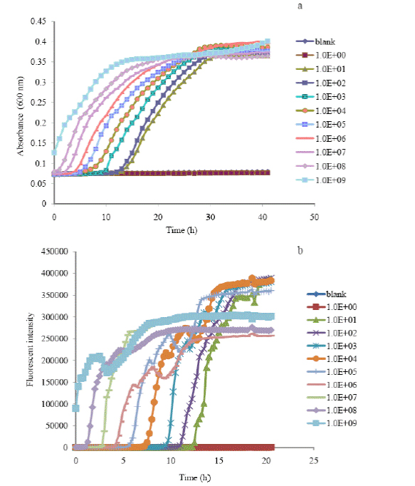
Figure 1. Time-growth profiles of absorbance (a) and oxygen flourenscence intensity (b) of E.
coli at different initial cell concentrations based on the turbidimetrec assay (a) and 
microrespirometic (b) assay. 
 
2. Rapid Detection of water- and foodborne pathogens by surface enhanced Raman spectroscopy
 
Surface enhanced Raman spectroscopy (SERS) is a novel and sensitive analytical tool that can be used in testing bacteria and virus samples using SERS-active substrates. Waterborne bacterial pathogens pose serious health risks to humans. Rapid, accurate, sensitive, and non-destructive detection methods are of great importance to protecting public health. This research task aimed to develop a new method to detect and discriminate E. coli O157:H7 and Staphylococcus epidermidis by SERS coupled with silver nanomaterials. An internal coating method using silver nanostructures for bacterial samples was established and assessed to achieve satisfactory SERS performance. SERS spectra were obtained from different bacterial cells (Figure 2). Distinctive differences in SERS spectral data between bacterial species were observed, specifically in the Raman shift region between 700-1500 cm-1 (Figure 2). The detection limit of SERS coupled with silver nanosubstrates could reach the level of single cells. Significant differences were observed between the spectra of E. coli O157:H7 and S. epidermidis. A mixture test for these two bacterial strains was also conducted (Figure 3). The results demonstrate that internal coating with silver nanostructures for bacterial cells is a feasible and effective approach to conduct SERS measurement for bacterial samples. Figure 4 shows the SEM pictures of S. epidermidis and E. coli O157:H7 cells internally coated with silver nanostructures. The results indicate that coupled with internal coated silver nanosubstrates, SERS was able to rapidly detect different water- and foodborne bacteria.
 
Figure 2
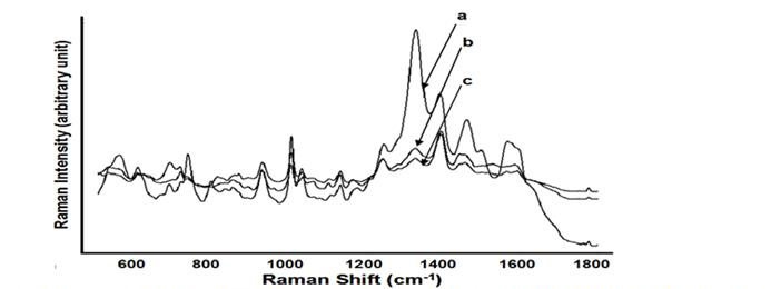
Figure 2. Average SERS spectra (n=7) acquired from 3 bacterial species with internal coating treatment
in a concentration of 109. s. epidermidis (a), E. coli K-12 (b), E. coli 0157:H7 (c).
 
 
Figure 3
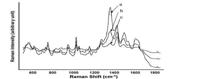
Figure 3. Average SERS spectra (n=7) acquired from S. epedermis, E. coli 0157:H7 and the 1:1 ratio
mixture with a concentration of 109: S. epdermidis (a), and the 1:1 ration mixture of S. epidermidis and
E. coli 0157:H7 (b), E. coli 0157:H7 (c).
 
 
Figure 4
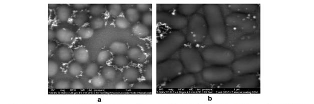
Figure 4. Scanning electron microscopy images of bacterial cells internally coated with silver 
nanostructures: S. epidermidis (a), E. coli 0157:H7 (b)
 
 
The SERS coupled with gold SERS-active substrates is also reliable for identifying and differentiating different virus strains. In this research task, SERS coupled with gold SERS-active substrates was used to detect and discriminate seven waterborne viruses, including norovirus- MNV4, adenovirus-MAD-1, parvovirus-MVM, rotavirus-SA-11, coronavirus-MVH, paramyxovirus-Sendai and herpersvirus-MCMV. These viruses can be classified into two categories: viruses with or without envelopes. By comparing the SERS spectral pattern of MNV4 obtained from a Klarite substrate with that from a flat good film substrate, the enhancement effects could be clearly visualized in Figure 5.
 
Figure 5
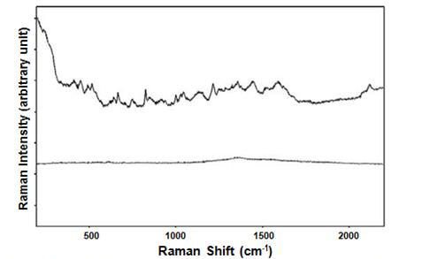
Figure 5. SERS spectrum and normal Raman spectrum of virus. 
 
Distinct differences in SERS spectra between different viruses were detected. Average SERS spectra of viruses acquired from eight virus strains was shown in Figure 6. All virus samples display fingerprint-like SERS spectra that are related to their unique structures. Soft independent modeling of class analogy (SIMCA) can differentiate the viruses with envelope and without envelope. Principle component analysis (PCA) shows clear spectral data segregations between virus strains with envelopes. The limit of detection of viruses with gold SERS-activate substrates could reach to a titer of 102. These results demonstrate that SERS method could provide rapid, sensitive, reproducible detection results with minimum sample preparation. Future work will include identifying and quantifying mixed waterborne pathogens using SERS.
 
Figure 6
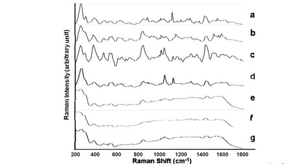
Figure 6. Average SERS spectru (n=5) acquired from 7 viruses with a titer ranging from 106 -107
in deionized water: Norovirus-MNV4 (a), Adenovirus-MAD-1 (mouse) (b), Parvovirus-MVM (mouse)
(c), Rotavirus-SA-11 (monkey)(d), Coronavirus-MVH (mouse) (e), Paramyxovirus-Sendai (mouse)
(f), Herpesvirus-MCMV (mouse) (g).
 
 
3. Enumeration of microbes using standard quantitative real-time PCR (qPCR)
 
Compared with traditional flocculants such as FeCl3 and Al2(SO4)3, chemical flocculation by lanthanum chloride (LaCl3) resulted in higher bacterial concentration and recovery efficiencies. The objective of this study was to develop and evaluate a lanthanum-based bacterial concentration method coupled with quantitative real-time PCR (qPCR) to enumerate bacteria in water. Here, we developed and evaluated a lanthanum-based concentration method coupled with quantitative real-time PCR to detect selected bacteria (E. coli and Helicobacter pylori) in water. To improve qPCR efficiency, the flocs with enmeshed bacteria after chemical flocculation need to be dissolved before PCR detection. Ethylenediaminetetraacetic acid (EDTA) salt successfully dissolved the flocs from a lanthanum-based flocculation process, but not those from the traditional processes using chemicals such as FeCl3 or Al2(SO4)3. Lanthanum-based concentration coupled with real-time PCR successfully determined E. coli at a concentration of 15 cells/mL in the raw and finished water and H. pylori at a concentration of about 1 cell/mL in the finished water prior to disinfection. By eliminating the membrane-clogging problem that is often encountered in direct membrane filtration, the lanthanum-based chemical flocculation coupled with qPCR is a promising method for concentration and determination of low density of microbial suspensions in water.
 
The protocols of qPCR reactions to determine E. coli concentrations were followed as described in the literature. PCR reactions which target the lacZ gene of E. coli were carried out in MicroAmp optical reaction plates containing 1 × TaqMan Universal Master Mix, 0.3 µmol/L of each primer, 0.2 µmol/L probe dual-labeled with a 5’-FAM (6-carboxyfluorescein) reporter dye and a 3’-TAMRA (6-carboxy-N,N,N’,N’-tetramethylrhodamine) quencher dye, and 5 µL of extracted DNA in a 25 µL total reaction volume. PCR reactions which target the ureA gene of H. pylori (McDaniels et al., 2005) contain 12.5 µL 2 × TaqMan Universal Master Mix, 1 µmol/L of each primer, 0.4 µmol/L probe, 0.2 mg/mL bovine serum albumin, and 5 µL of extracted DNA in a total of 25 µL reaction volume. Primers and probes used in this study can be seen in Table 1. For both PCRs, amplification was conducted at 50 °C for 2 min, followed by 10 min at 95 °C, 40 cycles of 95 °C for 15 s and a 60 °C annealing/extension step for 1 min in an ABI Prism 7500 Sequence Detection System (Applied Biosystems, CA).
 
Table 1. Primers and probes used for bacteria detection
Bacteria Target gene Amplicon size (bp) Primer and Probe Sequence (5'-3')
E. coli lacZ 180 Forward primer (lacZf)
CTTAATCGCCTTGCAGCACA
Reverse Primer (lacZr)
CAGTATCGGCCTACAGGAAGA
Probe (lac-ZTM)
ATTCGCCATTACAGGCTGCGCAA
h. pylori ureA 135 Forward primer (HpyF1)
GGGTATTGAAGCGATGTTTCCT
Reverse primer (HpyR1)
GCTTTTTTGCCTTCGTTGATAGT
Probe (HpyP1
AAACTCGTAACCGTGCATACCCCTATTGAG
 
The quantitative PCR assay coupled with lanthanum-based concentration was extended to the detection of low-density viruses in water. Direct membrane filtration is often used to concentrate viruses in water, but it may suffer from severe membrane fouling and clogging. In this research task, a lanthanum-based flocculation method coupled with modified membrane filtration procedures was developed and evaluated to detect viruses in large volume (40 L) water samples. The lanthanum-based flocculation method could easily reduce the water sample volume by a factor of 40. Additional volume reduction was achieved by a two-step membrane filtration approach. First, selected membrane filters (including 1MDS electropositive filters and nitrocellulose electronegative filters-Millipore HATF filters) were used to reduce water sample volume further and compare their efficiencies in virus recovery. The Mg2+-modified HATF membrane performed better on MS2 retention with an average virus recovery of 83.4% (± 4.5% [standard deviation]). More than 70% of mouse adenovirus (initial concentration = (8.01 ± 1.26) × 104 VP in 40 L or 2 VP/mL) could be concentrated after lanthanum coagulation/flocculation (Table 2). After floc dissolution and filtration, no virus was detected in the filtrate (data not shown), indicating all adenovirus particles were adsorbed on the modified HATF membrane.
 
 
Table 2. MOuse Adenovirus numbers in each step of the lanthanun-based concentration and membrane filtration procedures from the qPCR analycic
  Volume (mL) Viral particles recovered (± SD) Mean recovery (± SD) %
Lanthanum flocs 1000 5369 (± 0.62) x 104 70.7 (± 7.7) 
Filtrate 1000 ND ND
Eluate 15 8.35 (± 1.38) x 104 103.9 (± 17.2)
Centrifugation 2.35 5.51 (± 3.630) x 104 68.5 (± 44.8)

The total number of adenovirus spiked inot the 40-L decholrinated tap water (n=3) was 8.01 (± 11.26) x 104 VP
adn the initial virus concentrataion was 2 adenovirus particles/mL. ND = Not Detected. 


After HATF membrane filtration and elution, centrifugal ultrafiltration through a 30 k Dalton cut-off membrane resulted in an overall concentration factor of 20,000. Results from the infectivity assay showed that the MS2 recovery efficiencies from the NanoCeram- and 1MDS- based direct filtration and the lanthanum-based concentration coupled with the modified filtration procedure were 10.1% (± 1.0%), 3.3% (± 0.1%), and 17.5% (± 1.1%), respectively. Results from the PCR analysis showed the virus recovery of the lanthanum-based method at 20.6% (± 2.9%) and 19.5% (± 3.4%) for MS2 and adenovirus, respectively, while no adenovirus could be detected through the NanoCeram- and 1MDS-based direct filtration. The lanthanum- based concentration method coupled with modified membrane filtration procedures is therefore a promising method for detecting waterborne viruses.

Conclusions:

To summarize, a new method using lanthanum-based chemical flocculation was successfully developed for PCR detection of pathogens in water samples. EDTA completely dissolved the flocs from the lanthanum-based flocculation process, with additional bacterial concentration options through two-step flocculation, filtration and centrifugation. The lanthanum-based concentration method consistently showed higher virus detection efficiency than the currently EPA-approved methods in this study. No PCR inhibition was found throughout each concentration step. Furthermore, it took less than 4 hours with the lanthanum-based concentration/membrane filtration method compared to at least 6 hours required to complete all steps in the direct filtration method using Nanoceram or 1MDS. This project presents a proof of concept implementation of the pathogen concentration procedures, which could lead to new efforts to concentrate and detect low concentrations of pathogens in water with larger volumes and mixed microbial species.


Journal Articles on this Report : 5 Displayed | Download in RIS Format

Publications Views
Other project views: All 6 publications 5 publications in selected types All 5 journal articles
Publications
Type Citation Project Document Sources
Journal Article Fan C, Hu Z, Riley LK, Purdy GA, Mustapha A, Lin M. Detecting food-and waterborne viruses by surface-enhanced Raman spectroscopy. Journal of Food Science 2010;75(5):M302-M307. R833840 (2009)
R833840 (Final)
  • Abstract from PubMed
  • Abstract: Wiley Online-Abstract
    Exit
  • Journal Article Fan C, Hu Z, Mustapha A, Lin M. Rapid detection of food-and waterborne bacteria using surface-enhanced Raman spectroscopy coupled with silver nanosubstrates. Applied Microbiology and Biotechnology 2011;92(5):1053-1061. R833840 (2010)
    R833840 (2011)
    R833840 (Final)
  • Abstract from PubMed
  • Abstract: SpringerLink-Abstract
    Exit
  • Journal Article Zhang Y, Riley LK, Lin M, Hu Z. Lanthanum-based concentration and microrespirometric detection of microbes in water. Water Research 2010;44(11):3385-3392. R833840 (2009)
    R833840 (Final)
  • Abstract from PubMed
  • Abstract: ScienceDirect-Abstract
    Exit
  • Journal Article Zhang Y, Riley LK, Lin M, Hu Z. Determination of low-density Escherichia coli and Helicobacter pylori suspensions in water. Water Research 2012;46(7):2140-2148. R833840 (2010)
    R833840 (2011)
    R833840 (Final)
  • Abstract from PubMed
  • Full-text: ScienceDirect-Full Text HTML
    Exit
  • Other: ScienceDirect-PDF
    Exit
  • Journal Article Zhang Y, Riley LK, Lin M, Purdy GA, Hu Z. Development of a virus concentration method using lanthanum-based chemical flocculation coupled with modified membrane filtration procedures. Journal of Virological Methods 2013;190(1-2):41-48. R833840 (Final)
  • Abstract from PubMed
  • Abstract: ScienceDirect-Abstract
    Exit
  • Supplemental Keywords:

    Drinking water, exposure, waterborne pathogens, microbiology, monitoring, measurement methods, physical processes, health effects, field-based detection, lanthanum, respirometry, Surface Enhanced Raman Spectroscopy, nanotechnology, biotechnology, RFA, Scientific Discipline, Health, PHYSICAL ASPECTS, Water, Ecosystem Protection/Environmental Exposure & Risk, Health Risk Assessment, Environmental Chemistry, Monitoring/Modeling, Risk Assessments, Physical Processes, Environmental Monitoring, Drinking Water, microbial contamination, monitoring, measurement , microbial risk assessment, virulence factor biochip, virulence factor activity relationships, microbiological organisms, detection, exposure and effects, bacteria monitoring, exposure, other - risk assessment, E. Coli, human exposure, microbial risk management, microorganism, measurement, assessment technology, drinking water contaminants, other - risk management

    Progress and Final Reports:

    Original Abstract
  • 2008
  • 2009 Progress Report
  • 2010 Progress Report
  • 2011 Progress Report
  • Top of Page

    The perspectives, information and conclusions conveyed in research project abstracts, progress reports, final reports, journal abstracts and journal publications convey the viewpoints of the principal investigator and may not represent the views and policies of ORD and EPA. Conclusions drawn by the principal investigators have not been reviewed by the Agency.

    Project Research Results

    • 2011 Progress Report
    • 2010 Progress Report
    • 2009 Progress Report
    • 2008
    • Original Abstract
    6 publications for this project
    5 journal articles for this project

    Site Navigation

    • Grantee Research Project Results Home
    • Grantee Research Project Results Basic Search
    • Grantee Research Project Results Advanced Search
    • Grantee Research Project Results Fielded Search
    • Publication search
    • EPA Regional Search

    Related Information

    • Search Help
    • About our data collection
    • Research Grants
    • P3: Student Design Competition
    • Research Fellowships
    • Small Business Innovation Research (SBIR)
    Contact Us to ask a question, provide feedback, or report a problem.
    Last updated April 28, 2023
    United States Environmental Protection Agency

    Discover.

    • Accessibility
    • Budget & Performance
    • Contracting
    • EPA www Web Snapshot
    • Grants
    • No FEAR Act Data
    • Plain Writing
    • Privacy
    • Privacy and Security Notice

    Connect.

    • Data.gov
    • Inspector General
    • Jobs
    • Newsroom
    • Open Government
    • Regulations.gov
    • Subscribe
    • USA.gov
    • White House

    Ask.

    • Contact EPA
    • EPA Disclaimers
    • Hotlines
    • FOIA Requests
    • Frequent Questions

    Follow.