
Grantee Research Project Results
Final Report: Automated Methods for the Quantification and Infectivity of Human Noroviruses in Water
EPA Grant Number: R833831Title: Automated Methods for the Quantification and Infectivity of Human Noroviruses in Water
Investigators: Straub, Timothy M. , Ozanich, Richard , Bruckner-Lea, Cindy , Bartholomew, Rachel
Institution: Pacific Northwest National Laboratory
EPA Project Officer: Aja, Hayley
Project Period: July 1, 2008 through June 30, 2010 (Extended to June 30, 2011)
Project Amount: $592,140
RFA: Development and Evaluation of Innovative Approaches for the Quantitative Assessment of Pathogens and Cyanobacteria and Their Toxins in Drinking Water (2007) RFA Text | Recipients Lists
Research Category: Drinking Water , Water
Objective:
Optimization of water sample collection, sample processing, quantitative detection, and assessment of infectivity of human noroviruses in drinking water supplies will provide valuable occurrence information for both public health officials and the water industry.
Summary/Accomplishments (Outputs/Outcomes):
Monitoring of municipal water supplies (source and treated) for viral pathogens is a relatively labor intensive process that involves concentration of the viruses from 100-1,000 liters of water into a representative small (microliter (µL) to milliliter (mL)) volume that can be assessed by molecular methods such as real-time PCR (µL) and, for assessment of viral infectivity, tissue culture (mL). Concentration of viruses from large volumes to the small volumes required for analysis often co-concentrates inhibitors to PCR and/or compounds that are toxic to cell culture. Secondary concentration is required because volumes after primary concentration or elution of viruses from filters are too large, and inhibitors of PCR and cell culture are often present. At the purified small volumes suitable for detection, real-time PCR is gaining more popularity as well-designed assays can quantitate viral load. However, because PCR only detects nucleic acids that came from the viruses, it cannot be used as a measure for infectivity. Thus, cell culture is needed. However, for the human enteric viruses that can occur in water, several important strains cannot infect mammalian cell lines currently used for this purpose. This includes the human caliciviruses (norovirus) on the USEPA Contaminant Candidate List (CCL). From sample collection through analysis in cell culture as outlined in the draft USEPA Method 1615 for viruses in water, the process to confirm or rule out viral contamination of water can take weeks.
The ultimate goal of this project was to examine the process of waterborne virology monitoring and whether it could be optimized with regards to primary collection, secondary concentration to remove inhibitors and deliver purified samples suitable for quantitative PCR and cell culture, and whether these processes (with the exception of cell culture) could be automated. Our work was focused primarily on improved quantitative detection of human noroviruses in water and to apply innovative methods to assess human norovirus infectivity. Quantitative detection provides information on viral load that can be entered into quantitative microbial risk assessment (QMRA) models. Understanding whether the detected viruses are infectious provides important additional information for the QMRA to determine whether water requires further treatment or whether existing practices are suitable to remove risk. Below we summarize our findings in regards to the original specific aims our team proposed and how these findings are relevant for waterborne pathogen monitoring to protect public health.
Specific aim 1: Evaluate the automated concentration of viruses from large volumes of drinking water using hollow fiber ultrafiltration (HFF) in the context of a) engineering into an automated sampling system, and b) providing efficient and reproducible concentration of viruses within an automated system.
Currently, there is an expanding base of literature regarding methods for virus concentration from water, including the standard 1-MDS filter, the NanoCeram filter proposed for Method 1615, and hollow fiber ultrafiltration (HFF). Based on the literature, technical demonstrations our team attended, and laboratory investigations, our conclusions were that of the available options, dead end HFF was the best in terms of the potential to engineer the filtration system for automated collection. Molecular weight cutoff (MWCO) filtration using membranes that exclude particles less than 15,000 daltons will exclude viruses, bacteria, and protozoa parasites. These excluded "particles" remain in the retained volume after filtration (approximately 500 mL to 1 L), whereas the filtrate is discarded. The retained volume can be further processed to reduce volumes down to µL to mL volumes that are suitable for real-time PCR and/or tissue culture evaluation. Recovery efficiency for this method are often > 50% for viruses, bacteria, and protozoa, depending on the water sample.
Specific aim 2: Develop and validate fluidic secondary capture and purification of intact viruses from the retained volumes in HFF to allow analysis by PCR and cell culture.
Our team focused significant attention to this aspect of sample processing. Unlike USEPA Method 1623 for Giardia and Cryptosporidium, that has a relatively efficient immunomagentic separation step followed by an antibody dissociation step, similar methods do not exist for waterborne viral pathogens. In terms of automated sample processing, our team's approach was to investigate magnetic affinity reagents for efficient capture of norovirus. Due to the difficulty of working with human noroviruses, we started with bacteriophage MS2 as a surrogate virus to investigate issues involved with magnetic affinity reagents for the efficient capture of enteric viruses.
Dynabeads® Protein G magnetic beads were functionalized with antibodies to MS2 virus and tested both in rotational tube capture, to characterize antibody performance, and in our automated fluidics system. We chose to use Dynabeads® Protein G beads because of their potential to release virus by breaking the non-covalent bond between the bead and the antibody. This allows testing of captured target in cell culture for infectivity. In tube-based experiments, antibody capture, measured by disappearance of virus from supernatant was > 90%. In addition, the composition of filter elution buffer (HFF vs beef extract glycine used for viral elution from glass wool concentration) did not impact capture unless viral load exceeded 400 plaque forming units (pfu). When viral load was high, efficiency decreased, presumably due to saturation of the antibody binding sites. In terms of assessing infectious virus capture on the antibody, the team encountered several problems for which a solution was not apparent.
- Using a double agar overlay method to quantify capture, we applied this method to detect infectious MS2 captured on the beads. Without dissociation from the beads, the viruses were rendered "non-infectious." Only reverse-transcription real-time PCR could be employed to detect the captured virus. Moreover, a heat lysis step to free the viral RNA had to be employed as the magnetic beads caused inhibition of the real-time PCR
- The team was able to dissociate the Protein G-Ab complex from the magnetic beads. However, the team was unsuccessful in dissociating the virus from the Protein G-Ab complex, and was thus unable to restore viral infectivity. As an alternative, the team selected to utilize Dynabeads® M-280 Tosylactivated beads. These beads performed as well as the Protein G beads based on percent capture, but provide a covalent bond that would prevent loss of bound virus in fluidics.
Automated capture was the primary goal of this research, and the team developed a prototype system (Figure 1).
Figure 1: Prototype automated fluidics system for capture of enteric viruses from the retained volumes from primary HFF concentration.
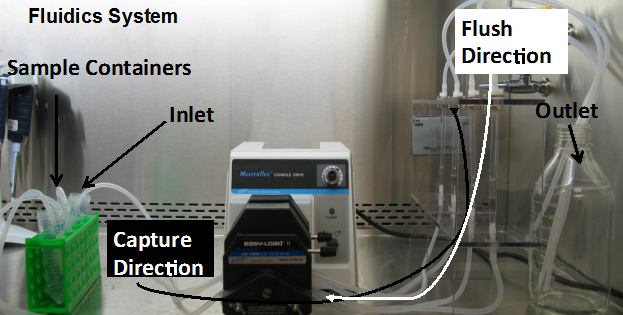
The automated system employs magnetic capture to concentrate the virus-Ab complex and purify it from other inhibitors in the system. Within the magnetized section to capture the beads, is a piece of nickel foam that is highly porous, paramagnetic, and captures the magnetic beads uniformly when the system is magnetized (as opposed to a wall based capture). This allows us to deliver a highly purified sample in a volume as small as 10 µL. This volume is ideal for real-time PCR detection. For an original water volume of 1,000 L, this represents a concentration factor of 108, and depending on viral load and sensitivity of the PCR should be representative of the water sample.
In the automated system, we tested capture of MS2 in HFF elution buffer and a beef extract elution buffer used for eluting viruses captured on glass wool. Only with the glass wool elution buffer was MS2 able to be recovered in the system. This problem was traced to loss of the virus to the nickel foam.
Work on the capture of human noroviruses was also investigated. We worked primarily with human norovirus GI.1 and antibodies to GI.1. Porcine gastric mucin (PGM) was also investigated as a less expensive alternative to antibody capture. The human norovirus capture experiments in batch proved more difficult than MS2 due to relatively poor efficiency of capture (approximately 10%). All work was based on comparison using real-time PCR. Without the benefit of a quantitative culture method like MS2, slight changes in Ct (cycle number that PCR was positive) had a dramatic effect on the actual quantity (determined by standard curve for each experiment). However, our general findings indicated that PGM performed equivalent to or, in some cases better than antibody capture. Like MS2, it was necessary to perform heat lysis to free the RNA from the magnetic beads. However, once in the supernatant (approximately 15 µL), quantitative reverse transcription real-time PCR could be performed. Internal positive controls for PCR, included with each experiment, showed that PGM capture followed by heat lysis provided samples that contained no PCR inhibition.
The findings from our automated fluidics work to capture viruses in concentrated water samples suggest that further optimization is needed to address:
- Efficient capture of the broad range of enteric viruses that may be present in a water sample. This includes the materials used in the fluidics architecture and buffers used in that system.
- Ability to dissociate the virus from the antibody complex so that infectivity studies can be conducted.
For now, the most efficient methods for secondary concentration appear to be either organic flocculation of beef extract (if beef extract is used as an eluting solution) or PEG precipitation. Neither of these methods is amenable to automated fluidics.
Specific aim 3: Optimize fast, quantitative reverse transcription real-time PCR (qRT real-time PCR) for the detection of the purified hNoV in drinking water.
We investigated several methods to improve the speed of qRT real-time PCR. Whereas there are a number of protocols that use one-step reverse transcription real-time PCR master mixes, we found that these improve speed to detection, but at a significant cost to efficient detection of viruses. Our recommendations to EPA are:
- Perform a separate reverse transcription reaction to convert viral RNA into cDNA. This separated step allows for analysis of different RNA enteric viruses because the cDNAs are generated from random, non-specific primers.
- For real-time PCR, include quantitative standards for each PCR plate run. These can be plasmid or RNA transcripts that have been converted back to cDNA. Use a master mix that contains dU instead of dT nucleotides, and incorporates uracil DNA glycosylase in the protocol to prevent PCR carryover contamination.
- For the noroviruses, use broadly reactive primer and probe sets that target GI and GII viruses, respectively. Use a master mix optimized for standard thermal cycling conditions (approximately 90 min per PCR).
Specific aim 4: Optimize our 3-D cell culture infectivity assay for hNoV, and test this assay for identification of infectious hNoV in spiked and unspiked drinking water samples.
The biggest success of this project was to identify factors leading to greater reproducibility of the norovirus infectivity assay. We published these results in the Journal of Water and Health, and the paper can be downloaded free from the following NCBI website: Human norovirus infection of caco-2 cells grown as a three-dimensional tissue structure
It is our hypothesis that due to the narrow tropism and specific binding requirements of human noroviruses to human intestinal epithelium that:
- Human cell lines derived from positive secretors must be used. We recommend the brush border-producing clone of Caco-2 cells.
- Brush border formation of the cells is required for infectivity. We have not determined whether this can be done as a transwell membrane assay or in the rotating wall vessel bioreactor configuration we described.
- Evidence of infectivity still needs to be assessed microscopically. Transmission electron micrographs are preferred to see evidence of microvilli blunting and cellular vacuolization. Real-time PCR can be used. In the most infectious samples, and at low dose, a 2-3 log increase in viral RNA titer can be observed.
Specific aim 5: Design and test process integration of HFF and secondary capture and purification, and evaluate the system performance. For the purposes of this project, the concentrated samples will be split such that half of the purified sample will be evaluated offline by qRT real-time PCR and the other half evaluated for infectivity in the 3-D cell culture
Given the challenges that we faced with optimization of the fludics aim in our research, we were unsuccessful in addressing this aim. As summarized above, the challenges will be efficient capture of a wide range of enteric viruses, and the ability to dissociate the virus from the affinity reagent so that infectivity studies can be performed.
Journal Articles on this Report : 1 Displayed | Download in RIS Format
| Other project views: | All 9 publications | 1 publications in selected types | All 1 journal articles |
|---|
| Type | Citation | ||
|---|---|---|---|
|
|
Straub TM, Bartholomew RA, Valdez CO, Valentine NB, Dohnalkova A, Ozanich RM, Bruckner-Lea CJ, Call DR. Human norovirus infection of Caco-2 cells grown as a three-dimensional tissue structure. Journal of Water and Health 2011;9(2):225-240. |
R833831 (Final) |
|
Supplemental Keywords:
Drinking water, microbial risk assessment, epidemiology, rotating wall vessel bioreactor, Biodetection Enabling Analyte Delivery System, waterborne pathogen viability, and quantitative reverse transcription real-time PCR;Relevant Websites:
Human norovirus infection of caco-2 cells grown as a three-dimensional tissue structure
Progress and Final Reports:
Original AbstractThe perspectives, information and conclusions conveyed in research project abstracts, progress reports, final reports, journal abstracts and journal publications convey the viewpoints of the principal investigator and may not represent the views and policies of ORD and EPA. Conclusions drawn by the principal investigators have not been reviewed by the Agency.