
Grantee Research Project Results
Final Report: Effects of Bisphenol A on the Developing Cortex
EPA Grant Number: R834593C004Subproject: this is subproject number 004 , established and managed by the Center Director under grant R834593
(EPA does not fund or establish subprojects; EPA awards and manages the overall grant for this center).
Center: Water Innovation Network for Sustainable Small Systems
Center Director: Reckhow, David A.
Title: Effects of Bisphenol A on the Developing Cortex
Investigators: Juraska, Janice
Institution: University of Illinois Urbana-Champaign
EPA Project Officer: Hahn, Intaek
Project Period: February 15, 2010 through February 14, 2014
RFA: Children's Environmental Health and Disease Prevention Research Centers: Formative Centers (with NIEHS) (2009) RFA Text | Recipients Lists
Research Category: Children's Health , Human Health
Objective:
The major goal of this Formative Center awarded in 2010 was to examine the effects of environmental chemicals on the developing neurological and reproductive systems. Project 4 was a pilot project designed to investigate whether bisphenol A (BPA) exposure during pre/perinatal development or during puberty alters cognitive areas of the brain or cognitive behavior. It modeled the effects of BPA in hooded rats, an animal model where sex differences in the cerebral cortex have been documented and are known to be influenced by the hormonal milieu during both the perinatal and peripubertal period. The effects of BPA on neuron number, a very basic building block of function, was explored in the medial prefrontal cortex, an area involved in higher functions in the mammalian brain including rats. Glia cells in the prefrontal cortex have been quantified as well. All quantification has been done with stereological methods. The behavioral consequences of cortical alterations also have been investigated in a visual spatial task, the radial arm maze, which shows sex differences in several laboratories.
Summary/Accomplishments (Outputs/Outcomes):
Four doses of BPA (0, 4, 40 or 400 µg/kg/day) were fed to male and female rats during early development (prenatally to dams and 10 days postnatally directly to the pups) or, in a separate experiment, during adolescence (days 27-46). Both cognitive behavior (17-arm radial maze) and cortical neuron number were quantified when the animals reached adulthood (>90 days). An addendum onto the original design is that preliminary observations of maternal behavior over the first 15 postnatal days were added to the early development experiment.
A. Pre/perinatal exposure to BPA
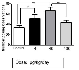
Maternal behavior. In one of the cohorts of the perinatal exposure groups, maternal behavior was monitored for the first 15 postnatal days. Some doses of BPA were found to increase maternal licking over controls. The group exposed to 40 µg/kg/day had significantly more licking than controls and the 4 µg/kg/day group also showed a trend toward more licking (Figure 1). The group exposed to 400 µg/kg/day did not exhibit more licking. No other behavioral category was affected by BPA in these dams. Thus, BPA exposure increased maternal attention to the pups at certain doses, which establishes that any effects of BPA are not due to insufficient maternal care. These data are preliminary at this time because of relatively low N. More animals will be added as part of the studies in the Center (P01) grant.
Figure 1. THe number of observations of maternal licking
of pups summed across 13 nights. Observations with night
vision goggles every 4 minutes per litter for 13 days.
** p,.0.2 *P<.04, #p=.07
The number of neurons and glia in the medial prefrontal cortex. The quantitative stereological analysis of the prefrontal cortex revealed that there were significant increases in the number of both neurons and glia in male rats exposed to 400 µg/kg/day of BPA perinatally compared to unexposed controls (Figure 2). No effects were found in exposed female rats. There are reports of an increase in the number of neurons and in the number of glial markers (astrocytes and microglia) in the prefrontal cortex of male children with autism (Courchesne, et al., 2011; Edmonson, et al., 2014), which may indicate that prenatal exposure to BPA is a predisposing factor for this syndrome.
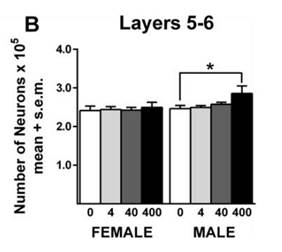
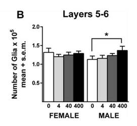
Figure 2. The number of neurons (left) and glia (right) in layers 5/6 of the perfrontal cortex. A similar pattern was found in the upper layers (2/3). Female rats
were unaffected on all measures.
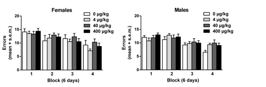
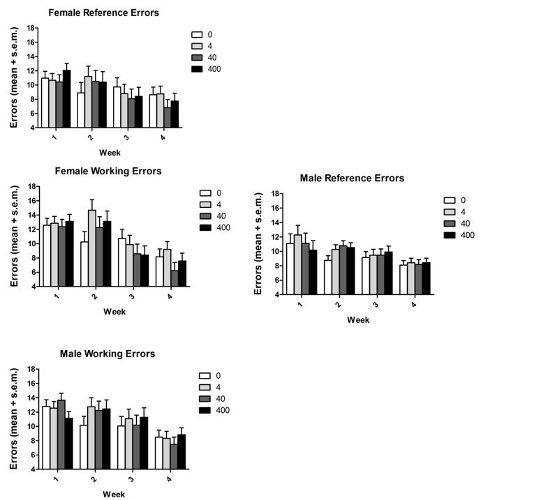
Performance on the baited/unbaited 17-arm radial maze. There were no significant changes in performance on the radial maze in animals exposed to BPA perinatally in either reference (long term) (Figure 3) or working (Figure 4) memory. It appears that this cognitive task does not reflect the effects of BPA on the number of neurons. Given the nature of the neural results, more emotional/social tasks might yield effects of pre/perinatal BPA exposure.
There also were no effects on body weight at weaning or in adulthood, or on levels of thyroid hormone or LH at weaning. Both sexes at doses of 4 and 40 µg/kg had decreased FSH levels but there were no changes in the timing of puberty. All of these results have been recently published (Sadowski, et al., 2014).
Figure 3. There were no differences due to BPA in reference memory errors (ams always balted) in eigher sex.
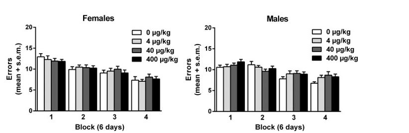
Figure 4. There were no differences due to BPA in working memory (arms never balted) in either sex
B. Pubertal exposure to BPA
Rats were exposed to BPA from postnatal day 27 through 45. As adults, they were tested on the 17-arm radial maze and then the number of neurons and glia were quantified in the medial prefrontal cortex.
Performance on the 17-arm radial maze. No effects due to BPA exposure were found (Figure 5).
Figure 5. There were no significant effects of exposure to BPA
during adolescense on performance on the 17-am radial maze.
Both reference and working memory erros are illustrated
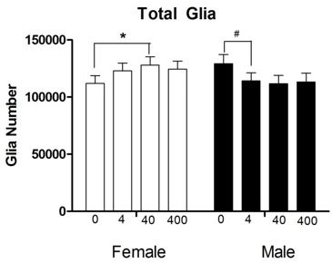
The number of neurons and glia in the medial prefrontal cortex. Exposure to BPA during adolescence did not change the number of neurons in the medial prefrontal cortex in either sex. However, glia were affected in opposite directions between the sexes with a near to significant sex by treatment interaction (p = .051). Post hoc comparisons between the vehicle and each dose for each sex revealed a significant increase in the number of glia between 0 and 40 µg/kg dose (p < .05) in females and a marginal decrease between 0 and 4 µg/kg (p < .06) in males (Figure 6). We currently are staining the extra sections from these brains for markers of two major types of glia: astrocytes and microglia. Both of these glia types are important for synaptogenesis and pruning, which may indicate an effect of BPA on the wiring of the medial prefrontal cortex.
Journal Articles:
No journal articles submitted with this report: View all 7 publications for this subprojectSupplemental Keywords:
Health, RFA, Scientific Discipline, Health Risk Assessment, Risk Assessments, Children's Health, Biochemistry, biological markers, children's vulnerablity, human health riskProgress and Final Reports:
Original AbstractMain Center Abstract and Reports:
R834593 Water Innovation Network for Sustainable Small Systems Subprojects under this Center: (EPA does not fund or establish subprojects; EPA awards and manages the overall grant for this center).
R834593C001 Prenatal Exposure to BPA/Phthalates: Infant Physical and Behavioral Development
R834593C002 Adolescent Exposure to BPA/Phthalates Cognitive and Behavioral Development
R834593C003 Mechanisms of In Utero BPA Exposure on Fetal Gonad Development
R834593C004 Effects of Bisphenol A on the Developing Cortex
The perspectives, information and conclusions conveyed in research project abstracts, progress reports, final reports, journal abstracts and journal publications convey the viewpoints of the principal investigator and may not represent the views and policies of ORD and EPA. Conclusions drawn by the principal investigators have not been reviewed by the Agency.